Japanese
English
- 有料閲覧
- Abstract 文献概要
- 1ページ目 Look Inside
Ⅰ.症例
患 者:Y. W. 66歳 家婦
主 訴:心窩部不快感
家族歴,既往歴:特記することなし.
初診:45年11月10日
手術施行日:45年12月20日
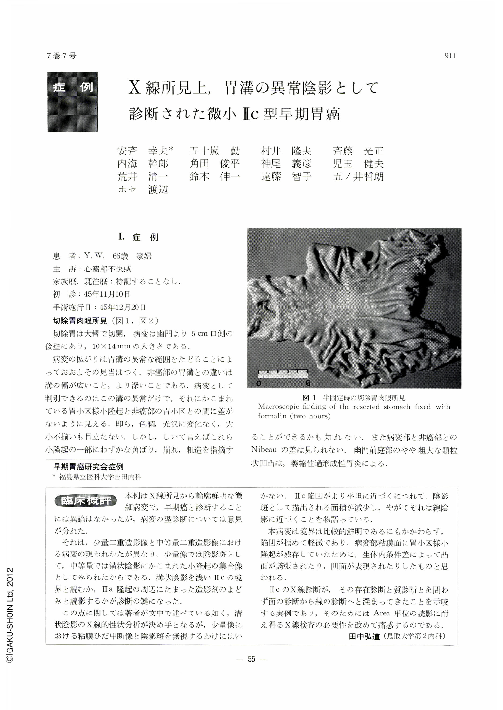
切除胃肉眼所見(図1,図2)
切除胃は大彎で切開,病変は幽門より5cm口側の後壁にあり,10×14mmの大きさである.
A very small lesion on the posterior wall of the antrum was demonstrated by x-ray as abnormal shadows of the gastric sulci, seen by endoscopy as linear reddening. It was then diagnosed as II c type early gastric cancer. Resected stomach revealed on the posterior wall 5 cm oral from the pylorus, a lesion measuring 10×14 mm, bordered by abnormal sulci. They were different from these of the non-cancerous areas in that they were wider and deeper. The only pathological finding in this case was the width of the sulci, and tiny area-like elevations surrounded by these sulci looking almost the same as those areas on the non-canerous parts. The height of the areae gastricae also showed no difference.
Histologically, the lesion proved to be adenocarcinoma mucocellulare with only m degree of depth invasion. Cancer was confined within the area surrounded by abnormal sulci. The only spot where cancer cells were exposed over the mucosal surface, was limited to the sulci between the areas, and the surface of the area-like elevations was covered with non-cancerous epithelia.
This tiny gastric cancer was characterized in x-ray as abnormal shadows of the sulci, and on that account the qualitative diagnosis of such a lesion as this, lies in the correct interpretation of these sulci. They are wider and more radiopaque, presenting fluffy and blurred shadows. Macroscopically, the lesion was flat, but by double contrast picture taken timely with smaller amount of air within the stomach, it could be demonstrated as if it were depressed. Compression is of no avail in such a tiny lesion. If located on the anterior wall, it may escape unnoticed by a routine x-ray examination.
As this case somewhat differs from typical patterns of II c hitherto shown both by x-ray and gross findings, we have reported it in detail with some discussions on its x-ray diagnosis.

Copyright © 1972, Igaku-Shoin Ltd. All rights reserved.


