Japanese
English
- 有料閲覧
- Abstract 文献概要
- 1ページ目 Look Inside
症例
患 者:菅○喜○,64歳,男.
主 訴:食思不振
家族歴:姉が胃癌で死亡.
既往歴:44歳の時胃潰瘍に罹患.
現病歴:昭和45年1月頃から心窩部膨満感が出現し某医にて胃レ線検査を受けたが異常無しと言われた.昭和46年3月頃から上記症状に加えて食思不振,体重減少も出現し,4月11日再び某医にて胃レ線検査を受けたところ異常陰影を指摘され,本学第1内科を紹介され,昨年4月22日外来を受診した.受診時持参した胃レ線所見から胃粘膜下腫瘍を疑い,当日直ちに胃内視鏡検査および胃生検を行ない,胃粘膜下腫瘍の診断のもとに5月7日本学第2外科に入院,5月11日胃亜全摘術を施行した.
入院時現症:体格中等度,栄養やや不良,結膜に貧血,黄疸無し.Virchowリンパ節を触れず,胸部に異常所見無し.腹部では心窩部に圧痛を認めた.
A 63-year-old man had a bout of dull pressure feeling in the epigastrium in January 1970. When he consulted his doctor, no change was found in the stomach by routine x-ray examination. In March 1971 he had in addition anorexia and weight loss, so that he visited his doctor again. At an x-ray examination done on April 11,1971, an abnormal shadow defect was found on the posterior wall of the antrum near the greater curvature. He was then referred to our hospital on April 22.
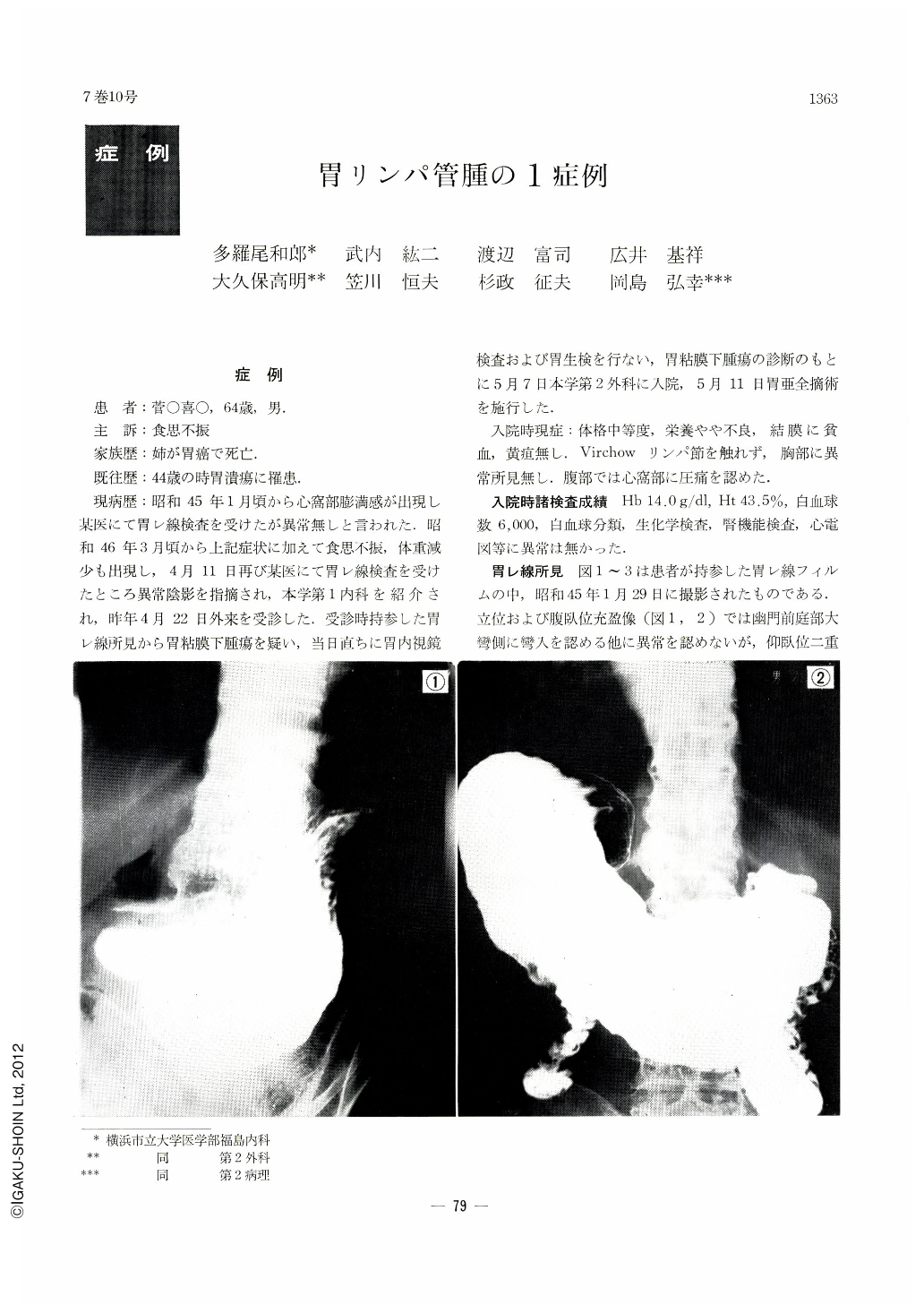
X-ray study of the stomach revealed an oval shadow defect the size of a quail's egg in double contrast picture on the posterior wall of the antrum in the greater curvature side. It was of smooth surface and accompanied with bridging folds. The filling defect varied in size and shape upon graded pressures. Endoscopy disclosed in the above area a hemispheric tumor with bridging folds over its margins. Resected stomach showed a rhomboid tumor, measuring 4.5×2.0×1.3 cm, located on the posterior wall of the antrum near the greater curvature. Bridging folds were seen over its margins. The cut surface of the tumor was of greenish-blue color. Subsequent histological examination revealed the tumor to be a lymphangioma dilated like a cyst.

Copyright © 1972, Igaku-Shoin Ltd. All rights reserved.


