Japanese
English
- 有料閲覧
- Abstract 文献概要
- 1ページ目 Look Inside
- サイト内被引用 Cited by
サルコイドージスは原因不明の全身性類上皮細胞肉芽腫症であり,縦隔および末梢リンパ節,肺,肝,脾,皮膚,眼,指骨,耳下腺その他の臓器を侵すが,胃に発生することは稀であり,欧米で30数例1),本邦で11例2)~11)の報告をみるにすぎない.
われわれは,直視下胃生検にて,確診にはいたらなかったが類上皮細胞肉芽腫を証明することのできた胃サルコイドージスの1例を経験したので報告する.
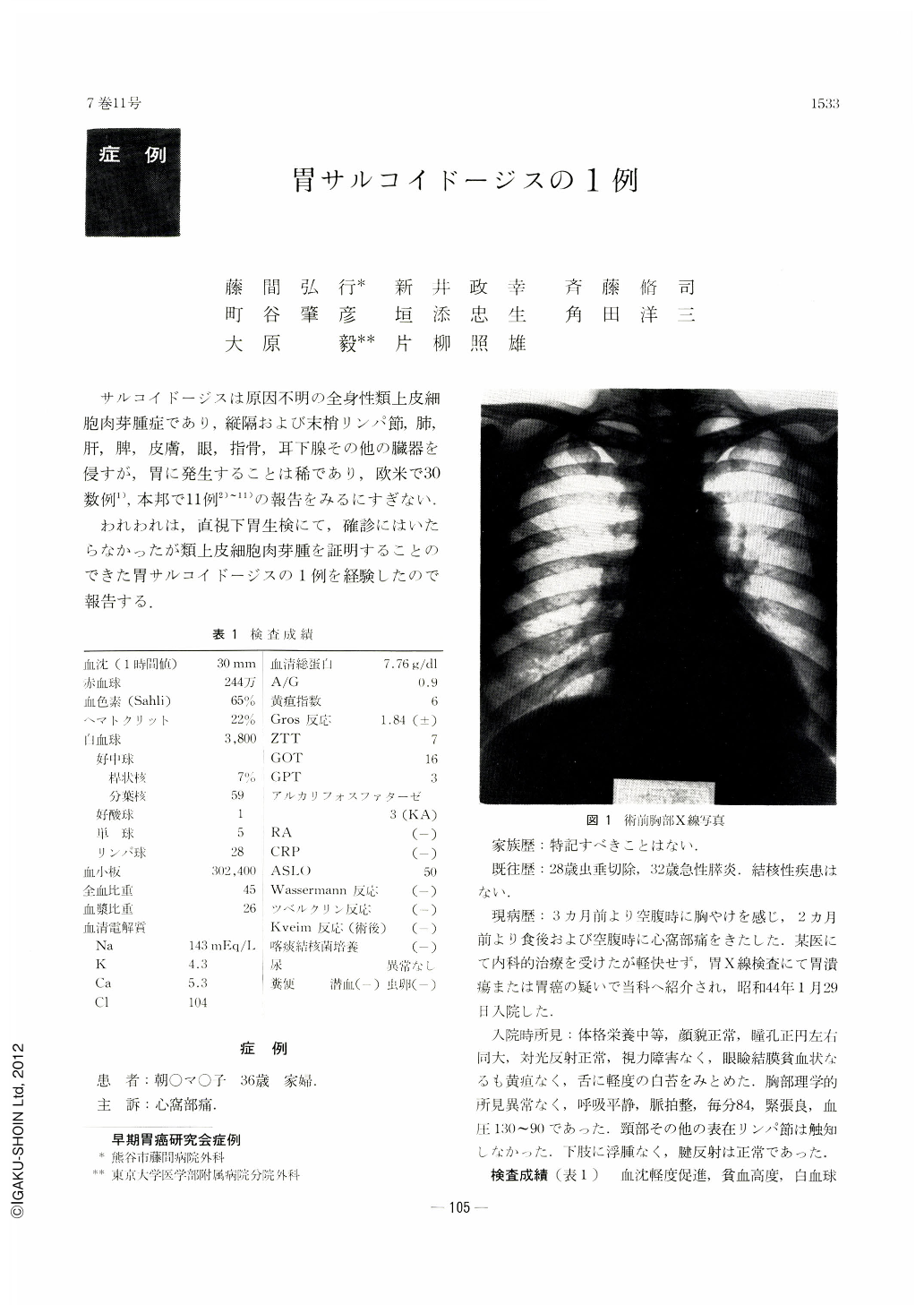
The patient is a 36-year-old housewife with a chief complaint of pain in the pit of the stomach of three months' duration. Laboratory examinations revealed anemia (65% sahli) ; both tuberculin and Kveim's reactions were negative. X-ray film of the chest was non-contributory. Roentgen study of the stomach showed not only enlarged gastric angle with irregular rigidity of the lesser curvature but also mesh-like barium flecks on the posterior wall, suggesting as a whole either ulcer scar or depressed type early cancer. Endoscopy demonstrated extensive area of erosion from the level of the angle down to the antrum with islet-like mucosal residues here and there over it. This finding was suggestive of either sarcoma or reactive lymphoreticular hyperplasia. Biopsy under direct vision still complicated the picture, and now gastric tuberculosis seemed to account for clusters of epithelioid cells and multinuclear giant cells found thereby. Finally gastric resection was done under a tentative diagnosis of gastric tuberculosis with a possibility of either sarcoma or Ⅱc-like lesion. Resected stomach showed an irregular reddened area, measuring 10×8 cm, extending from the angle down to the antrum. Slight ulcers were seen in this area. Histologically, there were hyperplasia of epithelioid cells and multiple small granulomas of almost uniform size consisting of Langhans type multinuclear giant cells, not only in the deeper strata of the mucosa but also in every layer from the submucosa to the subserosa.
No fusion was seen among granulomas, nor was there any caseous degeneration within them. Surrounding lymphocytic infiltration was slight. Subacute ulcers of Ul-Ⅱ degree were seen here and there. In all of regional lymph nodes excised were found similar granulomas. Stain for acid-fest rods was negative. Consequently, the present case was diagnosed as sarcoidosis of the stomach. The patient was in good health when seen two years later, but a x-ray film of the chest taken then showed a few round shadows in both sides of the lung. We were unable then to rule out sarcoidosis of the lung.
Copyright © 1972, Igaku-Shoin Ltd. All rights reserved.