Japanese
English
- 有料閲覧
- Abstract 文献概要
- 1ページ目 Look Inside
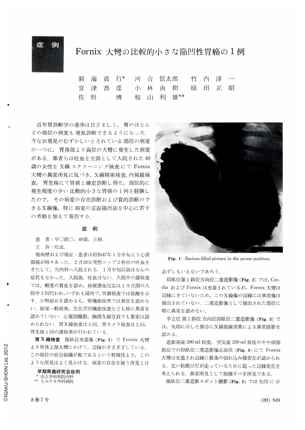
近年胃診断学の進歩は目ざましく,胃のほとんどの部位の病変も発見診断できるようになった.今なお発見のむずかしいとされている部位の病変の一つに,胃体部より高位の大彎に発生した病変がある.筆者らは吐血を主訴として入院された40歳の女性をX線スクリーニング検査にてFornix大彎の異常所見に気づき,X線精密検査,内視鏡検査,胃生検にて胃癌と確定診断し得た,部位的に発生頻度の少い比較的小さな胃癌の1例を経験したので,その病変の存在診断および質的診断のできるX線像,特に病変の正面描出法を中心に若干の考察を加えて報告する.
A report is made of a small carcinoma in the fornix of the stomach seen in a 40-year-old woman admitted to the hospital with a chief complaint of hematemesis. An abnormal finding detected on the greater curvature of the fornix by x-ray way was further scrutinized by detailed study combined with x-ray, endoscopy and biopsy, and a diagnosis of gastric cancer was established. Our description is centered around radiographs distinct enough to allow us to make not only existence diagnosis but also qualitative diagnosis of such a small carcinoma at a site least frequented by malignancy, with special reference to the most advantageous method to delineate such a lesion in en-face view.
The present case is an illustration of those cases for which even existence diagnosis of cancer was relatively difficult to achieve. In order to obtain an effective radiograph permitting us to make a qualitative diagnosis of cancer, we had to locate the lesion and to interprete to a certain degree its nature during fluoroscopy, and this attitude enabled us to take a good en-face view of the lesion. The present case has taught us how important good fluoroscopy is for exposing radiographs that permit correct interpretation.
The en-face view of cancer in this case was made in upright double contrast study in the first oblique position with the stomach over-distended with air.
Histological diagnosis was adenocarcinoma tubulare mucocellulonodulare.

Copyright © 1973, Igaku-Shoin Ltd. All rights reserved.


