Japanese
English
- 有料閲覧
- Abstract 文献概要
- 1ページ目 Look Inside
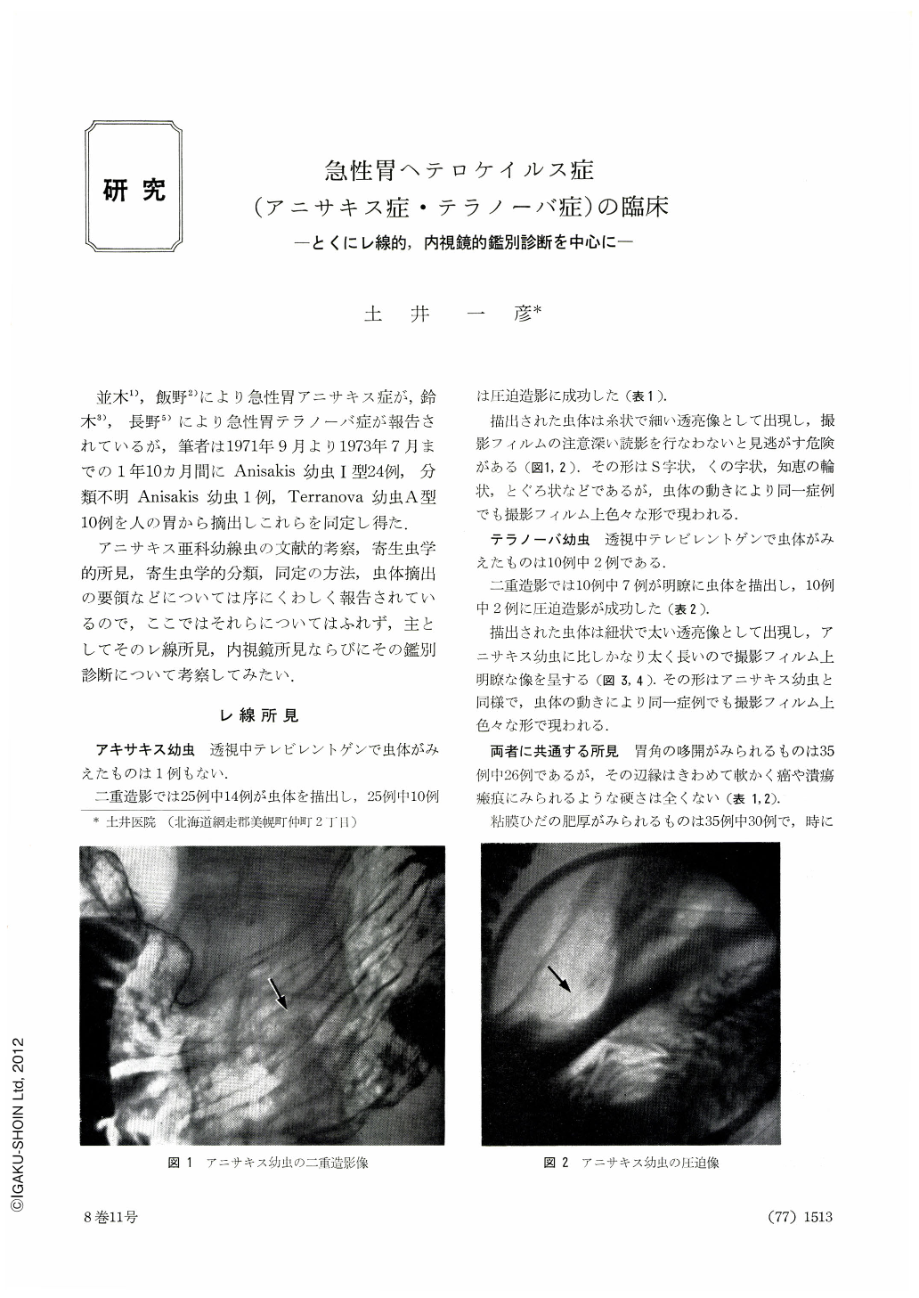
並木1),飯野2)により急性胃アニサキス症が,鈴木3),長野5)により急性胃テラノーバ症が報告されているが,筆者は1971年9月より1973年7月までの1年10カ月間にAnisakis幼虫1型24例,分類不明Anisakis幼虫1例,Terranova幼虫A型10例を人の胃から摘出しこれらを同定し得た.
アニサキス亜科幼線虫の文献的考察,寄生虫学的所見,寄生虫学的分類,同定の方法,虫体摘出の要領などについては序にくわしく報告されているので,ここではそれらについてはふれず,主としてそのレ線所見,内視鏡所見ならびにその鑑別診断について考察してみたい.
During the one year and ten months Sept. 1971 to July 1973 we have extracted anisakis larvae (type 1) out of the stomach in 25 cases and terranova larvae (type A) in 10. They were all identified.
Roentgenologically, anisakis larva is seen as a thin thread-like radiolucency, but unless utmost care is taken to delineate the details of the larva in double contrast study or compression picture, it can be easily overlooked. On the other hand, terranova larva is visualized as a thicker, string-like radiolucency, which can be pictured relatively easily in double contrast or compression view. Differentiation between the two is not so difficult.
When a suspicion of gastric larva is entertained either by the patient's history or his clinical picture, such x-ray findings as dilatation of the gastric angle or swollen mucosal folds provide us with a most important clue; even in cases where the body of the larva is not positively delineated in x-ray, the above-mentioned findings can be of great help in its detection by endoscopy.
In so far as endoscopic close-up view is concerned, the anisakis larva looks thin and milk-white, while that of the terranova is broader and of yellowish brown color. Differentiation between the two is thus possible.
The point at which the larva penetrated into the gastric wall and the surrounding mucosa are usually dotted with bleeding spots. The wall is often edematous and the mucosal folds, swollen. These can be useful landmarks to the detection of the larva at endoscopy.
In Bihoro Area acute gastric distress due to anisakis or terranova larva is caused in overwhelming numbers by eating raw Hippoglossus stenolepis. We are of the opinion that many cases in the past treated as acute gastritis must have been acute gastric heterocheilidiasis.

Copyright © 1973, Igaku-Shoin Ltd. All rights reserved.


