Japanese
English
- 有料閲覧
- Abstract 文献概要
- 1ページ目 Look Inside
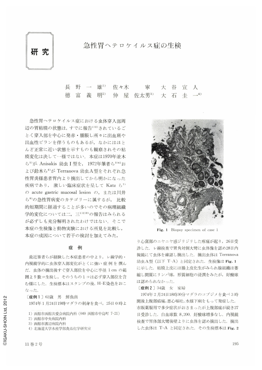
急性胃ヘテロケイルス症における虫体穿入部周辺の胃粘膜の状態は,すでに報告1)2)されているごとく穿入部を中心に発赤・腫脹し所々に出血班や出血性ビランを伴うものもあるが,なかにはほとんど正常に近い状態を示すものも観察されその粘膜変化は決して一様ではない.本症は1970年並木ら5)がAnisakis幼虫Ⅰ型を,1972年筆者ら3)4)および鈴木ら6)がTerranova幼虫A型をそれぞれ急性胃炎様患者胃内より摘出してから明かになった疾病であり,激しい臨床症状を呈してKatzら7)のacute gastric mucosal lesionの,または川井ら8)の急性胃病変のカテゴリーに属するが,比較的短期間に経過することが多いのでその病理組織学的変化については二,三1)2)9)の報告はみられるが必ずしも充分解明されたわけではない.そこで本症の生検像と動物実験における所見を比較し,本症の成因について若干の検討を加えてみた.
In acute gastric heterocheilidiasis, the gastric mucosa at the site and in the vicinity of larval penetration presents pathologic changes which, as previously reported, are on no account consistent but diverse in morphological features. This disease was first recognized in 1970 by Namiki et al. and the authors who discovered Anisakis larva type I and Terranova larva type A, respectively, removed them alive from the stomach of patients with symptoms indicative of acute gastritis. This disease belongs to the category of the acute gastric mucosal lesion described by Katz et al. or the acute gastric lesion of Kawai and his associates.
Because of the relatively brief course in most instances despite severe clinical symptoms, the histopathological findings in this disease have not been sufficiently elucidated although a few reports have been published. In view of this we studied the biopsy findings of the patient's stomach in comparison with the changes in laboratory animals with experimental gastric heterocheilidiasis, with the results providing the following suggestions of profound interest.
1) Biopsy in the lesions of larval penetration was performed on a series of seven patients with particularly marked findings demonstrable by x-ray and endoscopy. Biopsy disclosed virtually normal features of the mucosa or only changes common in already known non-specific gastritis in most of the cases, whereas in the few remainders were noted pathologic changes characterized by immediately subepithelial edema, fibrination, cellular (non-eosinophilic) infiltration and proliferation of lymph follicles.
2) These biopsy findings in the patients were assessed in comparison with the lesions of primary infection produced by the method of Oishi in rabbits by a single instillation with Terranova larvae type A isolated from cod, Gadus morrhua macrocephalus, and with the lesions of reinstillation in rabbits by six intermittent consecutive inoculations with Anisakis larve type I isolated from Theragra chalcogramma.
3) Some comments have been made on the pathogenesis of acute gastric heterocheilidiasis in the human, by deduction from the results 1) and 2).
Copyright © 1976, Igaku-Shoin Ltd. All rights reserved.