Japanese
English
- 有料閲覧
- Abstract 文献概要
- 1ページ目 Look Inside
- サイト内被引用 Cited by
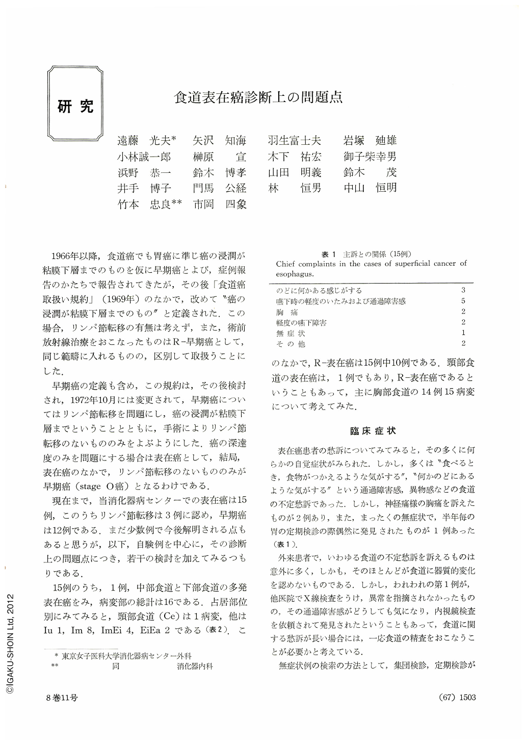
1966年以降,食道癌でも胃癌に準じ癌の浸潤が粘膜下層までのものを仮に早期癌とよび,症例報告のかたちで報告されてきたが,その後「食道癌取扱い規約」(1969年)のなかで,改めて“癌の浸潤が粘膜下層までのもの”と定義された.この場合,リンパ節転移の有無は考えず,また,術前放射線治療をおこなったものはR-早期癌として,同じ範疇に入れるものの,区別して取扱うことにした.
早期癌の定義も含め,この規約は,その後検討され,1972年10月には変更されて,早期癌についてはリンパ節転移を問題にし,癌の浸潤が粘膜下層までということとともに,手術によりリンパ節転移のないもののみをよぶようにした.癌の深達度のみを問題にする場合は表在癌として,結局,表在癌のなかで,リンパ節転移のないもののみが早期癌(stage 0癌)となるわけである.
From the clinical standpoint we have discussed some problems of superficial cancer of the esophagus localized within the mucosa or submucosa based on 14 cases with 15 lesions encountered by us. Undetermined complaints regarding the esophagus, if, they persist for a long time, should be checked by all means. It is desirable to examine the esophagus as well at mass screening or periodic check-up of the stomach. There was not even one case of esophageal cancer both x-ray and endoscopy found benign, but in about 33 per cent early cancer was mistaken for advanced one. A case of erosive depressed type reaching one fourth of the entire circumference was overlooked by x-ray. Such a lesion as this. only slightly uneven and falling short of the entire wall, should be visualized in a tangential direction. Endoscopy is more suitable for the diagnosis of such a case. Retrospective study of early cancer cases mistaken for advanced one shows that in tumor-like elevated type its size and shape looked exaggerated, as was also the case with marginal elevation in ulcer-like depression. Correct evaluation of the mural distensibility in the vicinity of the lesion is necessary along with more dynamic observation of the lesion itself. Pathologically, small lesions less than 2 cm in diameter were seen in more than half of our cases. Since such small lesions are reportedly found only in one per cent of advanced carcinoma, it is of great significance to look for such a small lesion in the diagnosis of early esophageal cancer. Comparision of endoscopic findings with the degree of cancer invasion shows that elevated type with its elevation hard to distinguish from the surrounding mucosa otherwise than by endoscopy and erosive depressed type were of mucosal cancer. Those in which the difference in height in resected specimens was more than 3 mm involved the submucosal layer without exception.

Copyright © 1973, Igaku-Shoin Ltd. All rights reserved.


