Japanese
English
- 有料閲覧
- Abstract 文献概要
- 1ページ目 Look Inside
患 者:今○光○ 68歳 男 用務員
家族歴:父74歳,黄疸にて死亡,母56歳,胃癌にて死亡.
既往歴:66歳時,心筋硬塞.
現病歴:昭和45年10月頃より体重減少,背部へ放散する上腹部痛より,次第に食思不振増強.昭和46年に入り,痛みが強まりEPGの結果,同年3月22日入院.経過中黄疸なく,貧血もなかった.
The patient: I. M., a 68-year-old man, school servant.
Familial history: At 74 his father had died of jaundice, and his mother, of gastric cancer.
Past history: At the age of 66 he had myocardial infarct.
Present history: Since about Oct. 1970 he began to lose weight and had pain in the upper abdomen radiating to the back. Anorexia became worse. Shortly thereafter in 1971 the pain became more intense. After examination with endoscopic pancreatography he was admitted to the hospital on March 22 (of the same year). Up to then no jaundice was noticed, nor was there any sign of anemia.
Blood chemistry at admission: Total protein, 7.4, A/G ratio, 0.8; glucose level, 127mg per 100ml; Al-P, 8 K. A. units; amylase level, 89s. units; icterus index, 6.
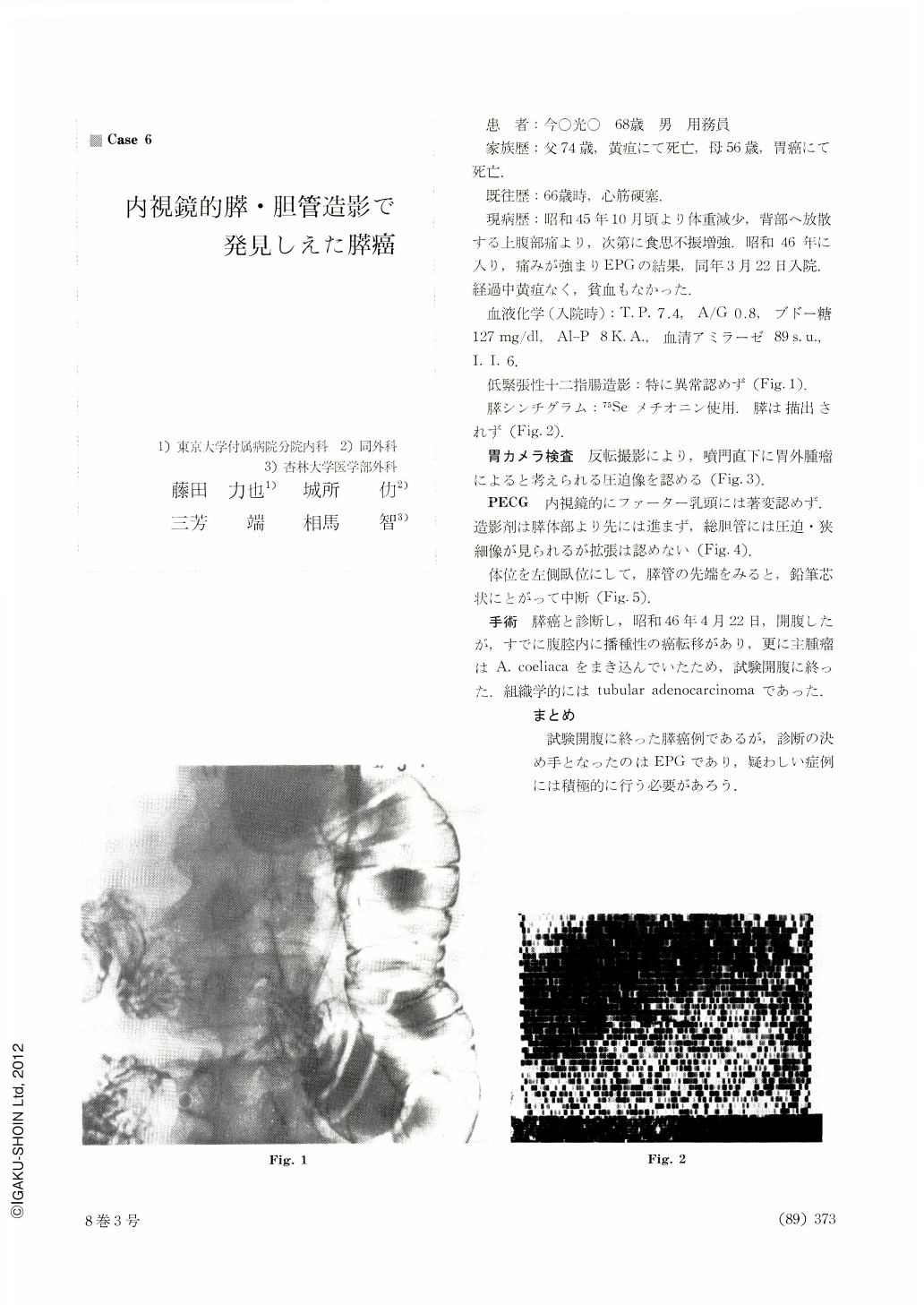
Hypotonic duodenography: non-contributory (Fig. 1).
Scintigram of the pancreas: 75Se methionine was used. The pancreas was not visualized (Fig. 2).
Gastrocamera examination: By reversed photography an abnormality, most probably due to extragastric tumor, was recognized at an area directly below the cardia corifice (Fig. 3).
Endoscopic Pancreatography: Endoscopically no particular change was seen in the papilla of Vater.
The contrast medium did not reach beyond the pancreatic body. The common bile duct was compressed and narrowed, and there was no dilatation (Fig. 4). A picture in the lateral position showed the tip of the pancreatic duct sharpened like a pencil point and then stopped up (Fig. 5).
Surgical operation: Under a diagnosis of pancreatic cancer laparotomy was done on April 22, 1971. Disseminated cancer metastases were seen within the abdominal cavity, with the main tumor involving A. coeliaca, so that the operation remained exploratory. Histologically cancer was tubular adenocaricinoma.
Comment: The most conclusive factor in the diagnosis of our case, although so advanced that the surgical operation remained after all exploratory, was endoscopic pancreatography and it should be positively employed whenever suspicious cases come up.
Copyright © 1973, Igaku-Shoin Ltd. All rights reserved.