Japanese
English
- 有料閲覧
- Abstract 文献概要
- 1ページ目 Look Inside
われわれは,Meckelの憩室を,術前に内視鏡的に診断し,潰瘍を確認しえた1例を経験した.これまで,Meckelの憩室の術前診断として小腸造影,血管撮影,99mTc‐pertechnetateを用いたシンチグラムなどが行われているが,内視鏡的に確認しえた報告はこれまでになく,これが世界で最初の症例と思われる.
症 例
患 者:斉○真○,7歳,男.
主 訴:下血.
家族歴,既往歴:特記すべきことなし.
現病歴:生来健康であったが,1978年6月18日,運動をして帰宅,午後4時ごろ腹痛出現したが,そのまま夕食は摂取した.午後8時ごろ,再び腹痛出現し下血.近医受診し止血剤投与され一時帰宅したが再び大量に下血し近医で輸液1,000mlを受け,翌19日当院小児外科に入院となる.
A case of Meckel's diverticulum with ulceration which was seen preoperatively by enteroscopy is reported. This is the first case ever seen by endoscopy so far.
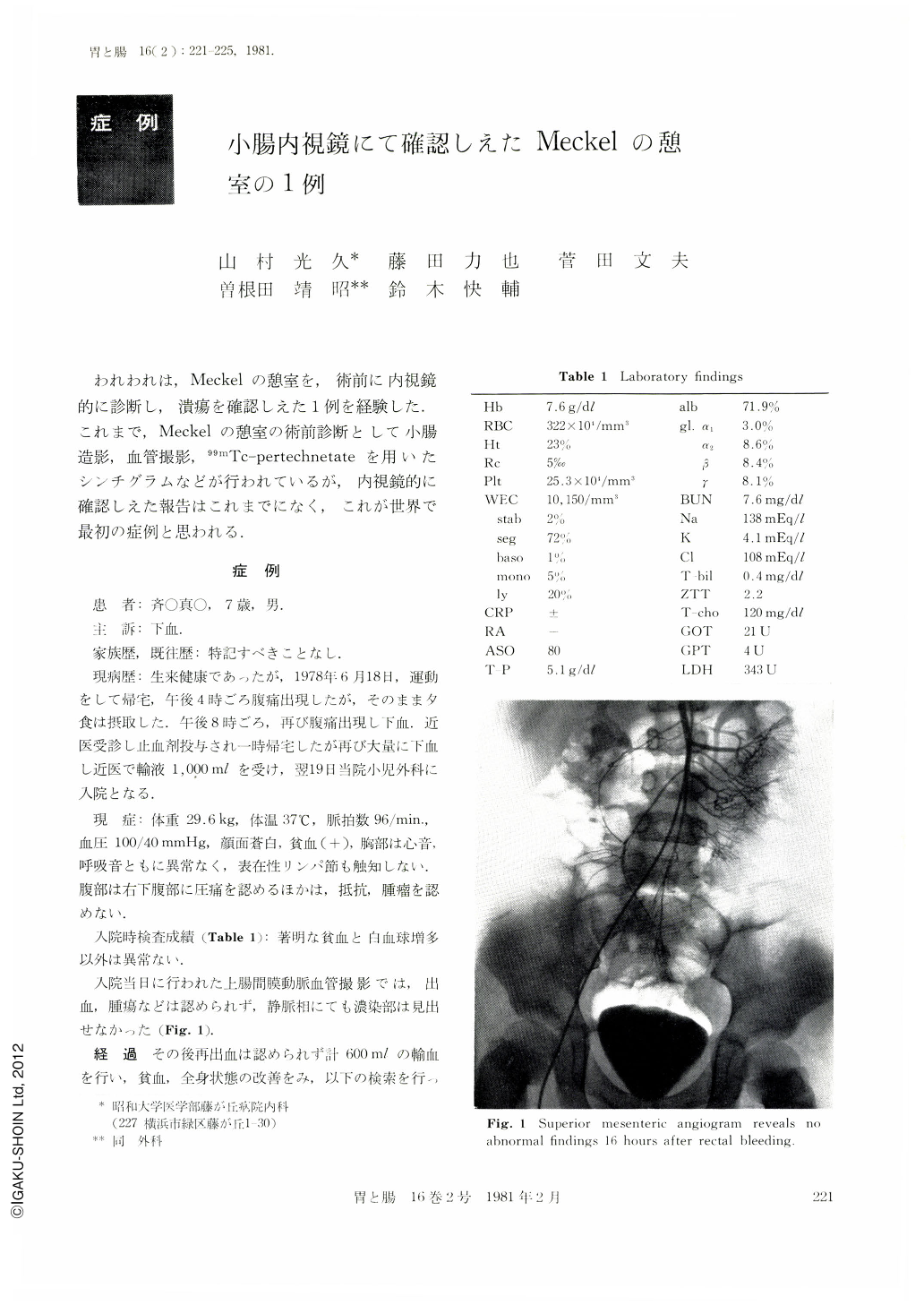
The patient is a 7 year-old boy who was admitted because of melena and anemia. Superior mesenteric angiogram was done firstly 16 hours after melena, but it showed no abnormal findings either in arterial phase or venous phase. Scintigram using 99mTc‐pertechnetate showed radioactivity accumulation in the right abdomen. Small bowel barium study showed the diverticulum by compression method. Retrograde enteroscopic procedure was performed according to the rope way method. Enteroscopy was made by long P2 (Olympus®, X-GIF-P2-2000), which has a special device; 750 mm longer than GIF‐P2. In this case, we were able to see the diverticulum and ulceration at about 45 cm from the terminal ileum. The diverticulum was resected by surgical operation and it demonstrated the ectopic gastric mucosa histologically.
Usually, barium study, angiography and 99mTc-scintigram are thought effective for the detection of Meckel's diverticulum.
In this report endoscopic observation of marginal ulceration at diverticulum is emphasized for the necessity of surgical indication, but it involves a complicated technique.

Copyright © 1981, Igaku-Shoin Ltd. All rights reserved.


