Japanese
English
- 有料閲覧
- Abstract 文献概要
- 1ページ目 Look Inside
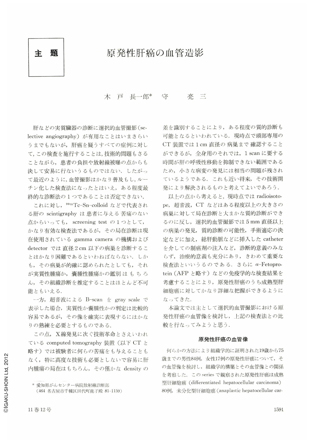
肝などの実質臓器の診断に選択的血管撮影(selective angiography)が有用なことはいまさらいうまでもないが,肝癌を疑うすべての症例に対して,この検査を施行することは,技術的問題もさることながら,患者の負担や放射線被曝の点からも決して安易に行ないうるものではない.したがって最近のように,血管撮影はかなり普及もし,ルーチン化した検査法になったとはいえ,ある程度最終的な診断法の1つであることは否定できない.
これに対し,99mTc-Sn-colloidなどで代表される肝のscintigraphyは患者に与える苦痛のない点からいっても,screening testの1つとして,かなり有効な検査法であるが,その局在診断は現在使用されているgamma cameraの機構およびdetectorでは直径2cm以下の病巣を診断することはかなり困難であるといわねばならない.しかも,その病巣が的確に認められたとしても,それが実質性腫瘍か,囊腫性腫瘍かの鑑別はもちろん,その組織診断を推定することはほとんど不可能ともいえる.
Eighty-four males and 17 females with histologically confirmed primary cancer of the liver ranging in age 19 to 75 years were studied for its vascular picture. Cholangioma was found in 15 cases and hepatoma in the remainder. Eighty cases belonged to the mature type and six to the anaplastic type.
In 94% of hepatoma of the mature type was noted vascular proliferation and in 72% of these cases, dilatation of abnormal vessels.
Tumor-stain is an important finding seen in 65%, in most of the cases appearing as nodular densities. Solitary tumors were generally round and their shape, size and position could be ascertained with considerable accuracy. When the tumor grows beyond a certain size or when it is growing rapidly, necrosis develops at the center and appears as a radiolucency in 15%.
Arteriovenous shunts are seen in 59%. While small shunts with intrahepatic portal vein occur in most cases (74%), shunts with the main trunk of the portal vein itself (13%) or with hepatic vein (13%) were also noted.
Proliferation of blood vessels, tumor-stain and formation of arteriovenous shunts represent the characteristics of mature type of hepatoma. In this tumor, tumor-cells are arranged in cords. There is little interstitial tissue and the cell cords are connected with endothelial cells, characterized by the presence of blood-filled sinusoids. Such a relationship between parenchymatous cells and sinusoids is exactly the same as that between normal hepatic parenchymatous cells and sinusoids. Liver is entirely filled with arterial blocd in hepatoma, so that the blood vessels are markedly dilated and tortuous and the contrast medium injected into the artery is retained for longer periods.
A-V shunts also occurs as the necessary consequence of such unusual hemodynamics. When the tumor enters the hepatic vein, forming tumor thrombus and causing an unequal distribution of blocd, various patterns of vascular pictures will emerge. Central necrosis produces avascular area in the arterial phase.
Proliferation of blood vessels was seen in 67% of hepatoma of the anaplastic type, but dilatation of pathological vessels was observed in only one case. In other cases, irregularities such as sclerosis were more conspicuous, with narrowing of the vessel lumen in many cases. Tumor-stain was observed in 33%; it was always diffuse and nonhomogenous and made the morphological definition of the tumor rather difficult. A-V shunt was seen in only one case. In the anaplastic type of hepatoma, the interstitial component became abundant, while blood distribution decreased, so that only hypervascularity of fine vessels and slight tumor-stain were noted.
In cholangioma, proliferation of blood vessels was noticed in 55%, but as in the anaplastic type of hepatoma, no dilatation was observed. Irregularity of the lumen was rather conspicuous. The proliferation was of the aboriation type; around the main trunk there was only proliferation of fine blood vessels. Obstruction and disruption of abnormal blood vessels was seen in 55%, and tumor-stain in 53%. In 4 of these cases, cyst-papillary adenocarcinoma probably arose from the main branch of the intrahepatic bile duct. In none of these cases was arteriovenous shunting observed. As specific findings, collateral circulation was noted in 20%. Tumor growth of cholangioma occurs obstructing the blood vessels, so that irregularity becomes conspicuous and proliferation of vessels is not pronounced. As the obstruction reaches the main trunk artery, other vessels dilate, playing the role of collateral vessels.
Comparison between the angiographic picture and the scintigram is an extremely difficult problem. The relationship between these methods was studied. Approximate agreement between the angiographic pattern and the hepatoscintigram with respect to the morphological aspects of the tumor such as its site, size and shape was observed in 50/68 (74%) of hepatoma of the mature type and 4/6 (67%) of anaplastic type. In cholangioma such an agreement was seen 6/15 (40%). The angiographic picture delineated the tumor more accurately than the scintigram in 14/68 (20%). The hepatoscintigram delineated the tumor better in 3/68 (4%) of mature type, 2/6 (33%) of anaplastic type, and 6/15 (40%) of cholangioma. In one case (2%) of hepatoma of mature type and in three cases of cholangioma, neither of these two methods provided us with useful information.
These facts seem to indicate that the angiographic findings are quite specific and interpretation is relatively easy in hepatoma of the mature type showing significant blood-vessel proliferation in both large and relatively small tumors. On the other hand, despite the presence of extensive changes of anaplastic hematoma and cholangioma, angiographic changes are not pronounced, and the information shown on the film may not be sufficient to make a diagnosis. The result shows a poorer diagnostic value of the angiographic picture as compared with the hepatoscintigram.
The hepatoscintigram was shown to be of great help not only as a screening test but also in the interpretation on the angiographic picture.

Copyright © 1976, Igaku-Shoin Ltd. All rights reserved.


