Japanese
English
- 有料閲覧
- Abstract 文献概要
- 1ページ目 Look Inside
われわれは最近,13年前に熊本大学第1外科でPeutz-Jeghers症候群と診断し,経過観察中に卵巣癌を合併して死亡した1例を経験した.本患者の多数の消化管ポリープのうち,回腸および結腸ポリープのおのおの1コに悪性化像が認められ,きわめて稀有な症例と思われるので報告する.
症 例
患 者:二○石○子 26歳 女性
主 訴:腹痛,嘔吐
既往症:幼児期から口唇の色素斑があった,1959年,9歳の頃から時々腹痛を訴えていた.1962年,12歳の時,腹痛を訴え,某医で慢性虫垂炎の診断の下に虫垂切除術を受けた.
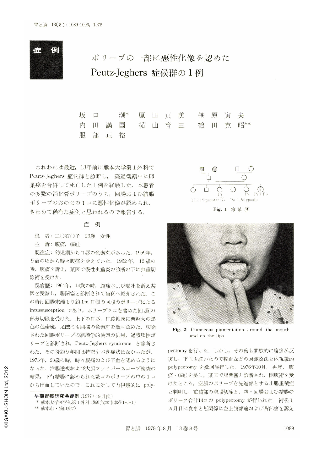
現病歴:1964年,14歳の時,腹痛および嘔吐を訴え某医を受診し,腸閉塞と診断されて当科へ紹介された.この時は回腸末端より約1m口側の回腸のポリープによるintussusceptionであり,ポリープ2コを含めた回腸の部分切除を受けた.上下の口唇,口腔粘膜に粟粒大の黒色の色素斑,足蹠にも同様の色素斑を数コ認めた.切除された回腸ポリープの組織学的検索の結果,過誤腫性ポリープと診断され,Peutz-Jeghers syndromeと診断された.その後約9年間は特記すべき症状はなかったが,1973年,23歳の時,時々腹痛および下血を認めるようになった.注腸透視および大腸ファイバースコープ検査の結果,下行結腸に認められた数コのポリープの中の1コから出血していたので,これに対して内視鏡的にpolypectomyを行った.しかし,その後も間歇的に腹痛が反復し,下血も続いたので輸血などの対症療法と内視鏡的polypectomyを数回施行した.1976年10月,再度,腹痛・嘔吐を呈し,某医で腸閉塞と診断され,開腹術を受けたところ,空腸のポリープを先進部とする小腸重積症と判明し,重積部の空腸切除と,空・回腸および結腸のポリープ合計14コのpolypectomyが行われた.術後1カ月目に食事と無関係に左上腹部痛および背部痛を訴えたので保存的治療を続けたが軽快しないので,1977年1月,熊本大学第1外科へ転医した.
The patient was a 26-year-old girl with pigmented spots on her lips and hands since childhood. Since nine years of age, she had occasional episodes of abdominal pain and was operated on for appendicitis at the age of 12.
At the age of 14 she was admitted to our hospital because of abdominal distension and vomiting. Examination revealed pigmented spots on the lips, buccal mucosa, palms and soles of feet. Roentgenographic examination of the abdomen demonstrated a niveau in the intestinal canal. Laparotomy was carried out with a clinical diagnosis of intussusception. A segment of the small intestine was resected and found to contain two polyps of a size of a walnut. Histologic diagnosis of the resected polyps was hamartomatous malformation. Her postoperative course was smooth and she was discharged with postoperative diagnosis of Peutz-Jeghers syndrome.
She was well until 1974, when she complained of lower abdominal pain and hematochezia. In 1975 she was admitted to the Tsuruta Hospital with anemia, and required a few times blood transfusion.
On barium enema study, about a dozen of polyps 2 to 4 cm in diameter with pedicles were seen distributed throughout the colon. Most of the polyps were removed via anus by endoscopical surgery, but intermittent hematochezia was still observed thereafter.
In 1976 she was readmitted to the Tsuruta Hospital because of recurrent abdominal pain with vomiting. Laparotomy was performed for intussusception of the jejunum due to a polyp.
A segment of the jejunum 10 cm long including the polyp was resected. In adition seven small bowel polyps and seven colonic polyps were removed by enterotomy and colotomy. Histologic features of the intestinal polyps were all hamartomatous polyps, and an atypical area interpreted as carcinoma in situ was found in two polyps; one in the jejunum and the other in the distending colon.
Three month months after operation on Jan. 10, 1977, she was readmited to our hospital because of left epigastralgia and back pain. Ascites and metastastic cervical lymph nodes were noticed three months later. The patient died at the age of 26 in May 1977. Autopsy revealed gastrointestinal polyposis distributed from the stomach to sigmoid colon, ovarian cancer with its' metastases to the pancreas and retroperitoneal lymph nodes and a great amount of ascites.

Copyright © 1978, Igaku-Shoin Ltd. All rights reserved.


