Japanese
English
- 有料閲覧
- Abstract 文献概要
- 1ページ目 Look Inside
最近のX線検査,内視鏡検査技術の進歩によって微小胃癌はかなり診断されるようになってきた.しかし胃上部,特に食道胃接合部の微小癌は,この部位の診断上の困難さもあり,発見されることは稀である.最近われわれは胸部X線写真を手がかりとして,食道胃接合部の胃平滑筋腫を発見し,更に内視鏡,および生検にてその表面粘膜上に微小Ⅱc型早期胃癌の併存することを術前に診断することが可能であった症例を経験したので報告する.
症 例
患 者:M. Y. 59歳 女性
主 訴:胸痛
家族歴・既往歴:特記すべきことなし
現病歴:10年前より時々胸痛あり,最近は1日3,4回の胸痛発作があった.1975年3月6日当院外科受診.
The patient was a 59-year-old woman with a chief complaint of chest pain.
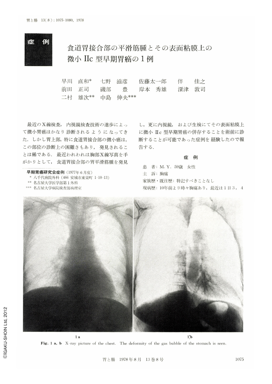
The gastric lesion was suspected with the chest X-ray examination at her first visit to our hospital. Subsequent barium meal X-ray examination revealed a submucosal tumor at the cardiac region of the stomach. An irregular-shaped red spot was found with endoscopy on the gastric anterior side of the tumor close to its central pit. The biopsy specimen from this spot was found to be adenocarcinoma. Laparotomy performed on March 11, 1975, showed a tumor close to the greater curvature at the esophago-gastric junction. The size of this tumor was 35 X 20 X 15 mm and a shallow depression, 5 X 15 mm in diameters, was found on it. Microscopic examination of this tumor revealed leiomyoma and that of the depression tubular adenocarcinoma, which spread under the esophageal mucosa.
Although 10 cases of minute early gastric cancer at the esophago-gastric junction have been reported, all were of elevated type. Our case is the first of depressed type. Furthermore, the presence itself of early gastric cancer on leiomyoma is rare.
While we failed to diagnose this case as a gastric cancer with X-ray examination, endoscopy and subsequent biopsy was useful. The endoscopic examination could be superior to the X-ray examination when the gastric lesion is extremely small.

Copyright © 1978, Igaku-Shoin Ltd. All rights reserved.


