Japanese
English
- 有料閲覧
- Abstract 文献概要
- 1ページ目 Look Inside
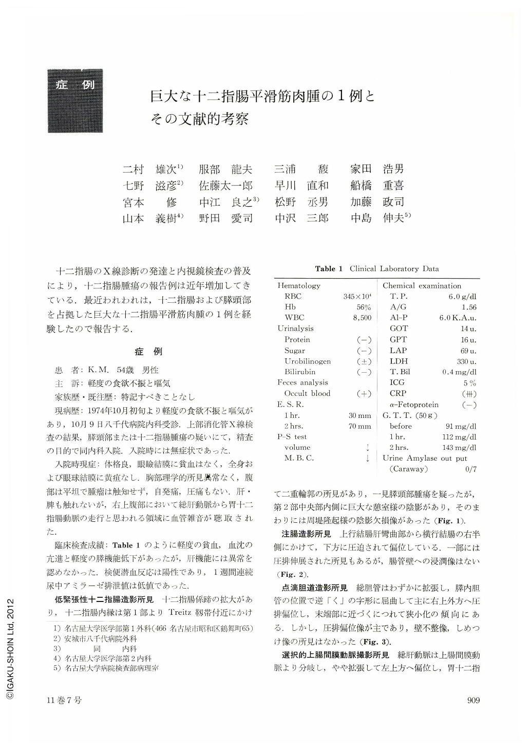
十二指腸のX線診断の発達と内視鏡検査の普及により,十二指腸腫瘍の報告例は近年増加してきている.最近われわれは,十二指腸および膵頭部を占拠した巨大な十二指腸平滑筋肉腫の1例を経験したので報告する.
A 54 year old man was seen on October 9, 1974, with complaints of slight anorexia and nausea. The pancreatic head or duodenal tumor was suspected from an X-ray examination of the upper G-I tract. A diagnosis of a malignant tumor of the duodenum was made from hypotonic duodenography, barium enema, drip infusion cholangiography, superior mesenteric arteriography, and endoscopic retrograde cholangiopancreatography. Duodenofiberscopy also revealed a large ulceration at the anal side of Papilla Vateri and a few biopsied specimens were taken from the lesion. Histological diagnosis was leiomyosarcoma.
On November 15, 1974, the operation was performed. We noticed a liver metastasis in the right hepatic lobe and a lymphnode metastasis in the mesenteric root, and performed panceaticoduodenectomy, cholecystectomy, right hemicolectomy, partial jejunectomy and partial right hepatectomy. In the resected specimens, the tumor, 12×11×11.5 cm in size, was situated at the anal side of Papilla Vateri and had an exenteric growth toward the pancreatic head and body.
Leiomyosarcoma of the duodenum is an uncommon malignant tumor and the first case was reported by Von Salis in 1920. McBrien reported 95 cases in 1971. In our country,84 cases were reported from 1939 to 1974. We have referred to the literature of leiomyosarcoma of the duodenum.

Copyright © 1976, Igaku-Shoin Ltd. All rights reserved.


