Japanese
English
- 有料閲覧
- Abstract 文献概要
- 1ページ目 Look Inside
近年胃X線,内視鏡検査は著しく進歩し,その形態学的診断能の向上に伴い多発性早期胃癌症例も数多く報告されるようになってきた.最近,われわれも術前にⅠ型とⅡc型多発早期胃癌と診断し,手術にてともに深達度sm,一部ブルンネル腺への浸潤を認めた症例を経験したので報告する.
症 例
患 者:由○治○郎 56歳 男性
主 訴:全身の紅斑
既往歴:20年前より糖尿病で治療をうけている.1977年6月16日,白内障(右)の手術.
家族歴:特記すべきことなし
現病歴:約6年前より顔面に瘙痒感を伴う紅斑と鱗屑が発生し色素沈着を残すようになった.その後本症状は増悪と寛解を繰返していたが,1977年1月頃より全身倦怠感も出現し,奈良医大付属病院皮膚科に入院した.
A report is made of a case of double early cancer of the stomach.
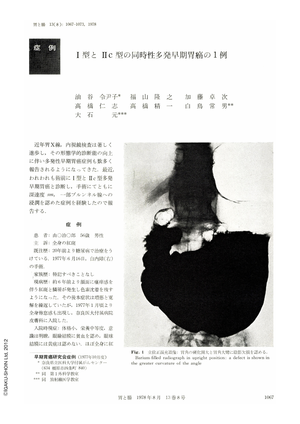
The patient, a 56-year-old man, was referred because of erythroderma of six years' duration. He was admitted to hospital because he had increasing general fatigue. At the time of admission, the patient was in moderate nutritional condition with average physique. Two lymphnodes in the cervical area were palpated with no lymphnode in the supraclavicular area. The abdomen, flat and non-resistant, had no trace of tangible tumor. The erythrocyte count in peripheral blood was 3.16 million per cu. mm., and Hb was 8.1 g/dl. Tests for urine remained within normal limits. Liver function tests showed that cholinesterase was 0.4 ΔpH; total protein was 5.1 g/dl. Immunoglobulins, IgG, IgA and IgM, were 2230 mg/dl, 472 mg/dl and 88 mg/dl. Occult blood in the feces was positive. Hypertrophy of the left heart ventricle and ischemic ST-T changes were seen in electrocardiogram. X-ray and endoscopic examination of the stomach revealed two lesions, a protruding lesion and a depressed one. The protruding lesion in the posterior wall near the greater curvature of the angle, clearly demarcated from the surrounding area, belonged to Yamada's Ⅲ type and was nodulary on the cardiac side. The surface was irregular and slightly reddened. The size was 20 X 40 mm. The depressed lesion with no concentration of mucosal folds was seen in the posterior wall near the lesser curvature of the antrum. Its margin was clear. The surrounding mucosa was slightly elevated and nodular. The size was 13 X 28 mm. Histologically the former was tubular adenocarcinoma of well differentiated type, INF-β and sm. The latter was tubular adenocarcinoma of moderately differentiated type, INF-β and sm. Invasion of gastric cancer cells into the region of the Brunner glands was seen.
Copyright © 1978, Igaku-Shoin Ltd. All rights reserved.