Japanese
English
- 有料閲覧
PPV購入案内
1,320円 (1,200円+税10%) こちらは、433ページから437ページの文献です。購入には医書.jp本会員登録が必要です。
会員登録完了後に改めて上記「購入サイトへ」を選択してください。
または「購入サイトへ」選択後に「購入ログイン」、「新規会員登録」の順に進んでください。
会員登録について詳しくはをご確認ください。
- Abstract 文献概要
- 1ページ目 Look Inside
回腸潰瘍より下血を来したBehçet病不全型と思われる症例を経験したので報告する.
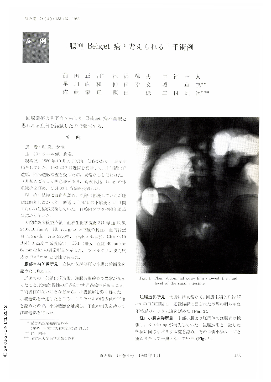
A 52 year-old woman visited our hospital complaining of abdominal distension, tarry stool and weight loss. Aphta in the oral cavity and/or genital ulceration were not found. A plain abdominal x-ray film showed the fluid level in the small intestine. An intestinal tumor was suspected because of the history of slowly progressive passage disturbance for six months and also because of no past history of laparotomy. Barium enema study showed the clearly-shaped barium spot surrounded with the elevated lesion at the ileum 17 cm from the Bauhin's valve.
Double contrast study of the small intestine showed the disapperance of Kerckring and the dilatation of the bowel. Selective ileo-colic angiography showed no particular findings and malignant lymphoma was suspected. 50 cm ileum including the tumor at the ileum end and the spleen were resected. Macroscopically, the main lesion was the deep ulceration 30×70 mm in size, and some ulcers were found at the oral and anal site. Microscopically, the ulcer was Ul-Ⅳ with a severe inflammatory cell infiltration and diagnosed as simple ulcer. Re-examination of the past history revealed that she had occasionally developed aphthous stomatitis for 15 years and suffered genital ulcer eight years ago. Postoperative course was good. However, erythema nodosum-like eruption on the right lower leg and aphthous stomatitis developed during the following six months.
Intestinal Behçet cannot be differentiated from simple ulcer either macroscopically or microscopically. Further observation is needed to clarify the nature of the disease.
Copyright © 1983, Igaku-Shoin Ltd. All rights reserved.