Japanese
English
- 有料閲覧
- Abstract 文献概要
- 1ページ目 Look Inside
われわれは,小腸のCrohn病として切除後4年目に残存小腸に再発し,回腸・回腸間の内瘻形成が認められたために再手術した症例を経験したので報告する.
症例
患 者:27歳 男
主 訴:腹痛
家族歴:特記することなし
既往歴:<20歳>1969年3月,臍周囲に痛みを訴え,時々嘔吐するようになった.その約2カ月後,急に強い腹痛が現れたので某医を受診し,イレウスの診断をうけ,手術の目的で某病院に入院した.諸検査の結果は異常が認められず,腹痛も軽快したため10目間で退院した.
<21~22歳> 時々軽い腹痛を訴えていたが放置していた.
<23歳>1972年9月4日,強い腹痛のために某医を受診し,再びイレウスの診断をうけ,精査治療の目的で9月6日に当センターへ入院した.入院後も強い腹痛が持続するため,イレウスの診断のもとに開腹術を施行した.
A 23-year-old-male was admitted to our hospital in Sept. 1972, because of severe abdominal pain. Diagnosed as small intestinal obstruction, immediate emergency operation was recomended. Then macroscopical and histological diagnoses were probably Crohn's disease of small intestine.
He was readmitted to our hospital in Sept. 1976, because of abdominal pain, abdominal fullness, anorexia, diarrhea, anemia and fever.
X-ray examination of small intestine with double contrast method showed skip lesions and longitudinal ulcer with pseudodiverticula in jejunum and internal fistulae and longitudinal ulcer in ileum. There was no remarkable change in large intestine by barium enema. Partial jejunoileotomy was again performed, diagnosed as Crohn's disease. Jejunum was resected about 25 cm in length and ileum, about 45 cm in length.
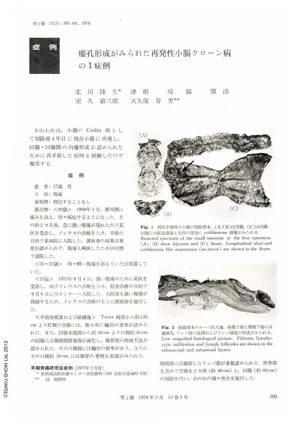
Macroscopical findings showed three small ulcers and longitudinal ulcer in jejunum and internal fistulae and longitudinal ulcer in ileum. All of these ulcers were located just beneath the attachment of the mesentery. Histological findings showed intramural inflammation, sarcoid-like granuloma in the subserosal layer and fissuring ulcers. From these findings, Crohn's disease of small intestine pattern was diagnosed.

Copyright © 1978, Igaku-Shoin Ltd. All rights reserved.


