Japanese
English
- 有料閲覧
- Abstract 文献概要
- 1ページ目 Look Inside
胃上部癌,特に噴門部癌は,最近のX線検査や11)~13)16)23)内視鏡検査技術の進歩にもかかわらず4)15)18)21),早期胃癌として発見される頻度が低い.最近われわれはいわゆる食道胃粘膜接合部のⅡa型早期癌を経験したので報告する.
In spite of recent progress in diagnostic techniques of radiology and endoscopy, cancer of the cardia is rarely seen in early phase, and the majority have been detected in advanced stage.
We have reported two cases of early cancer at the esophage-cardiac junction of the cardia.
Case 1
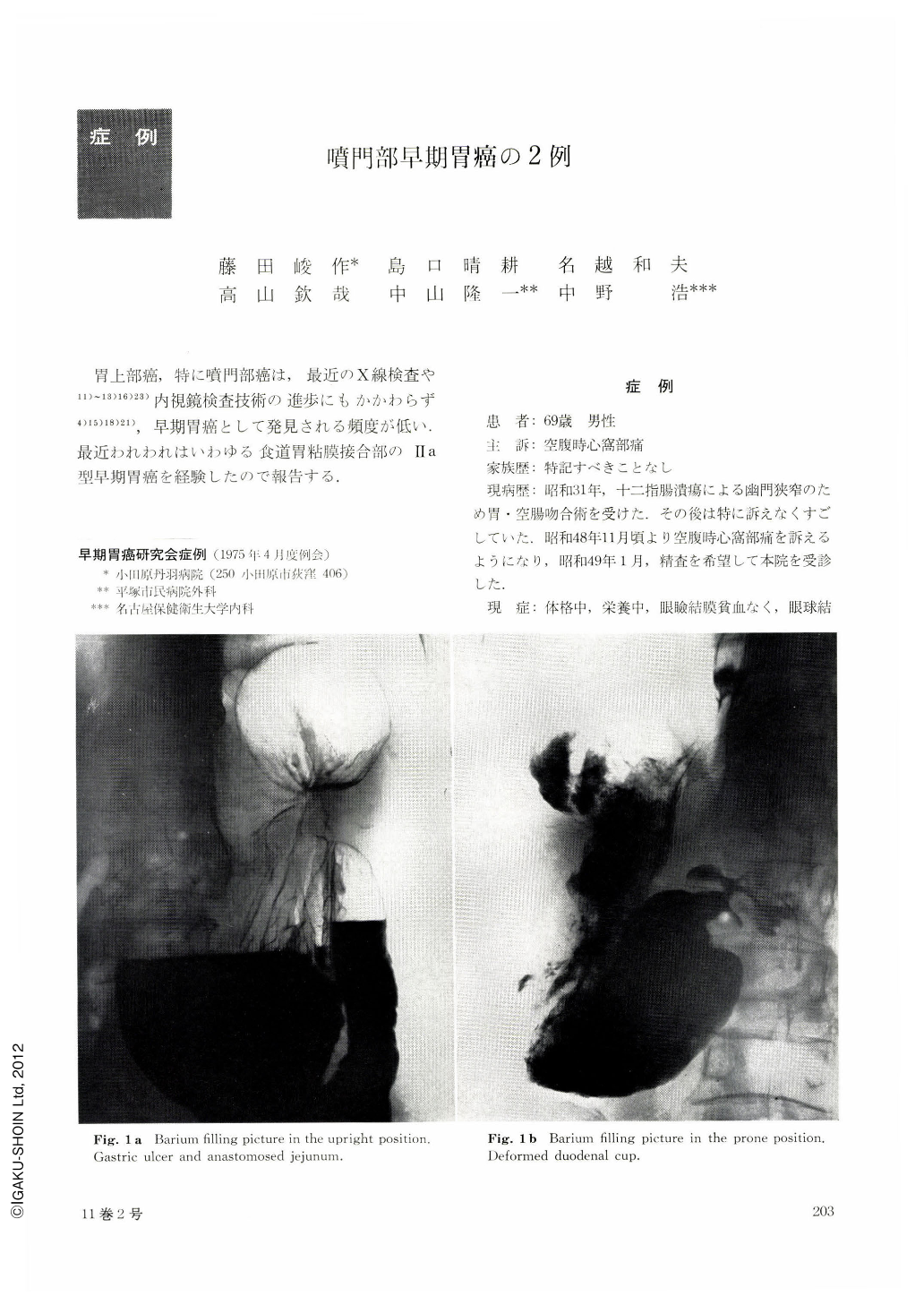
A 69-year-old man visited our hospital complaining of hunger pain. Roentgenographic examination of upper GI-tract revealed a gastric ulcer at the greater curvature, deformed duodenal cap and the jejunum anastomosed to greater curvature. Besides, a minute granular lesion was also disclosed at the cardia. Esophagofiberscopy found it a discolored and slightly elevated minute polypoid lesion at the cardia, and a few biopsy specimen were taken from it. Histological diagnosis was adenocarcinoma. Esophagogastrectomy was performed. In the resected stomach, a linear ulcer was seen at the greater curvature of gastric body, and slightly elevated, somewhat discolored lesion(histologically mucinous adenocarcinoma, type Ⅱa) measuring 12×12 mm was situated at the esophago-cardiac junction The infiltration of the cancer was limited within submucosal tissue.
Case 2
The man aged 63 had been complaining of epigastric distress. Roentgenographic and esophagoscopical examination of cardia showed the same finding as in case 1. In the resected specimen, slightly elevated carcinoma (histologically tubular adenocarcinoma, type Ⅱa) measuring 15×10 mm was seen at esophago-cardiac junction, limited within the submucosa. The disclosure of early cancer at the esophagocardiac junction is very rare. We have reported two cases of it, and briefly discussed about the roentgenographic skill to pick up the lesion.

Copyright © 1976, Igaku-Shoin Ltd. All rights reserved.


