Japanese
English
- 有料閲覧
- Abstract 文献概要
- 1ページ目 Look Inside
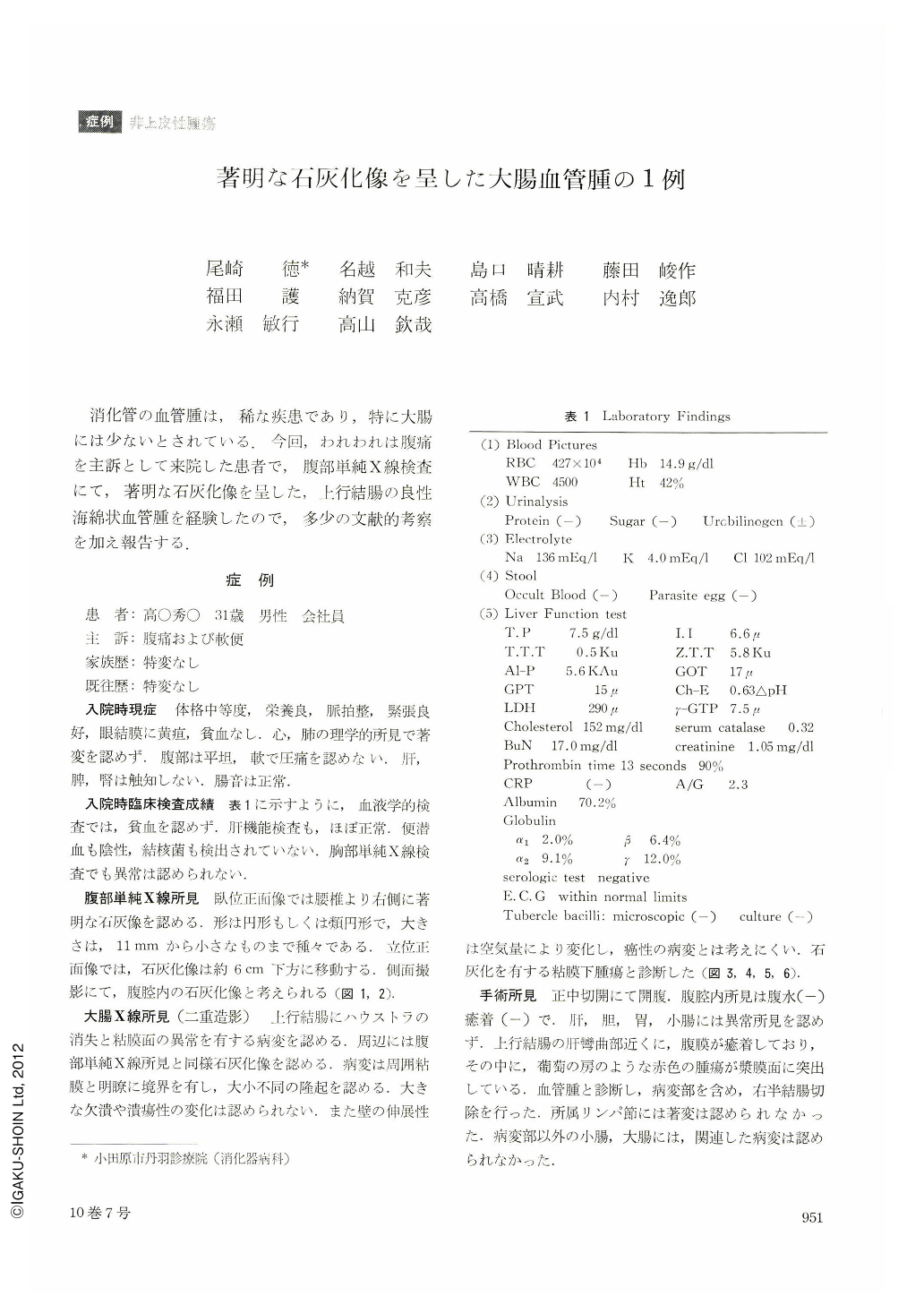
消化管の血管腫は,稀な疾患であり,特に大腸には少ないとされている.今回,われわれは腹痛を主訴として来院した患者で,腹部単純X線検査にて,著明な石灰化像を呈した,上行結腸の良性海綿状血管腫を経験したので,多少の文献的考察を加え報告する.
A man 31 years of age visited our hospital with chief complaints of abdominal pain and loose passages. Plain x-ray film of the stomach revealed nearly round calcifications in the right flank, at most 11 mm in the greatest diameter. X-ray examination of the colon showed disapearance of the haustrae with mural irregularity about 8 cm long in the ascending colon near the hepatic flexure. Calcifications were seen in the neighboring areas. The lesion was distensible and its picture varied according to the amount of air sent in. Laparotomy was done under a tentative diagnosis of submucosal tumor of the ascending colon with calcifications. The findings at operation corresponded well with those of x-ray. A cluster of tumors resembling a bunch of grapes in the ascending colon protruded from the serosal surface. As hemangioma was a most likely diagnosis, right hemicolonectomy was performed. The neoplasm was located 6 cm anal from the ileocecal valve, 8 by 8 cm in dimensions, of dark red color with surface irregularities. In the center were seen erosive changes. The cut surface showed grayish white calcifications within the dilated vessels.
Histologically celluar atypicality was scarce and the tumor proved to be benign hemangioma cavernosum.

Copyright © 1975, Igaku-Shoin Ltd. All rights reserved.


