Japanese
English
- 有料閲覧
- Abstract 文献概要
- 1ページ目 Look Inside
- サイト内被引用 Cited by
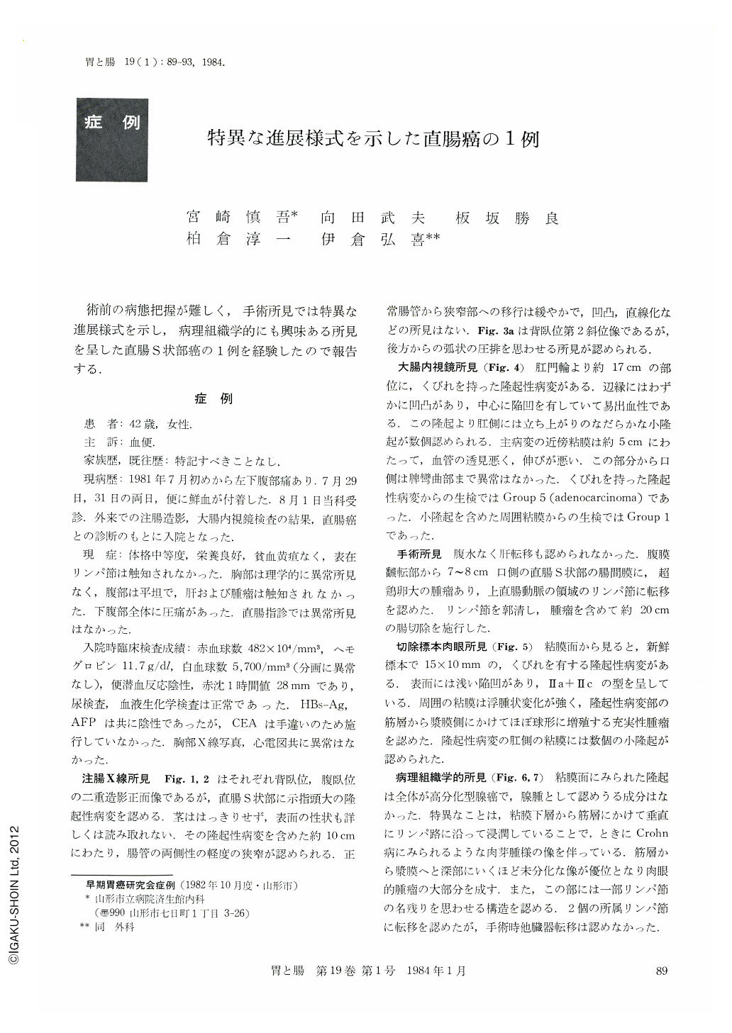
術前の病態把握が難しく,手術所見では特異な進展様式を示し,病理組織学的にも興味ある所見を呈した直腸S状部癌の1例を経験したので報告する.
The patient, a 42 year-old woman, visited our hospital with a chief complaint of bloody stools. Barium enema examination showed in the rectosigmoid colon a protuberant lesion and slight stricture in its neighborhood. Colonofiberscopic examination revealed in the same place edematous change of the wall and poor distension. At a place 17 cm oral from the anal ring was seen a protruding lesion strictured at the base with central depression. Histological study of the biopsy specimens showed adenocarcinoma, but otherwise no malignant finding was seen in the adjacent edematous mucosa.
The rectosigmoid mucosa of the resected specimen was edematous with a tumor mass formation, measuring 15×10 mm. Underneath the protrusion was seen another tumor far bigger than a hen egg. Histologically the mucosa was highly edematous and the tumor seen in the mucosa was a mass of well-differentiated adenocarcinoma with no vestige of adenoma. Cancer infiltrated vertically along the lymphatic vessels from the submucosa to muscle layer. As it reached deeper from the muscle layer to serosa poorly differentiated cancer cells became more dominant. In some parts were seen structures suggesting traces of lymph nodes. Metastasis was seen in two regional lymph nodes, but at the time of operation other organs were not involved. The present case was advanced colon cancer showing small growth in the mucosa but unusual development forming an extramurally large tumor.
Histogenesis of colon cancer has often been discussed such as adenoma-carcinoma sequence and de novo cancer. The present case seemed to suggest the latter.

Copyright © 1984, Igaku-Shoin Ltd. All rights reserved.


