Japanese
English
- 有料閲覧
- Abstract 文献概要
- 1ページ目 Look Inside
- サイト内被引用 Cited by
患者:57歳,男性
主訴:心窩部痛,家族歴に特記すべきことなし
既往歴:48歳胆囊結石にて,胆剔術を受く
現病歴:1976年4月(術前1年10月)より心窩部痛を訴え,痛みは断続的で時に疝痛様であって,1年間痛みが続き,1977年5月24日本院内科を受信す.
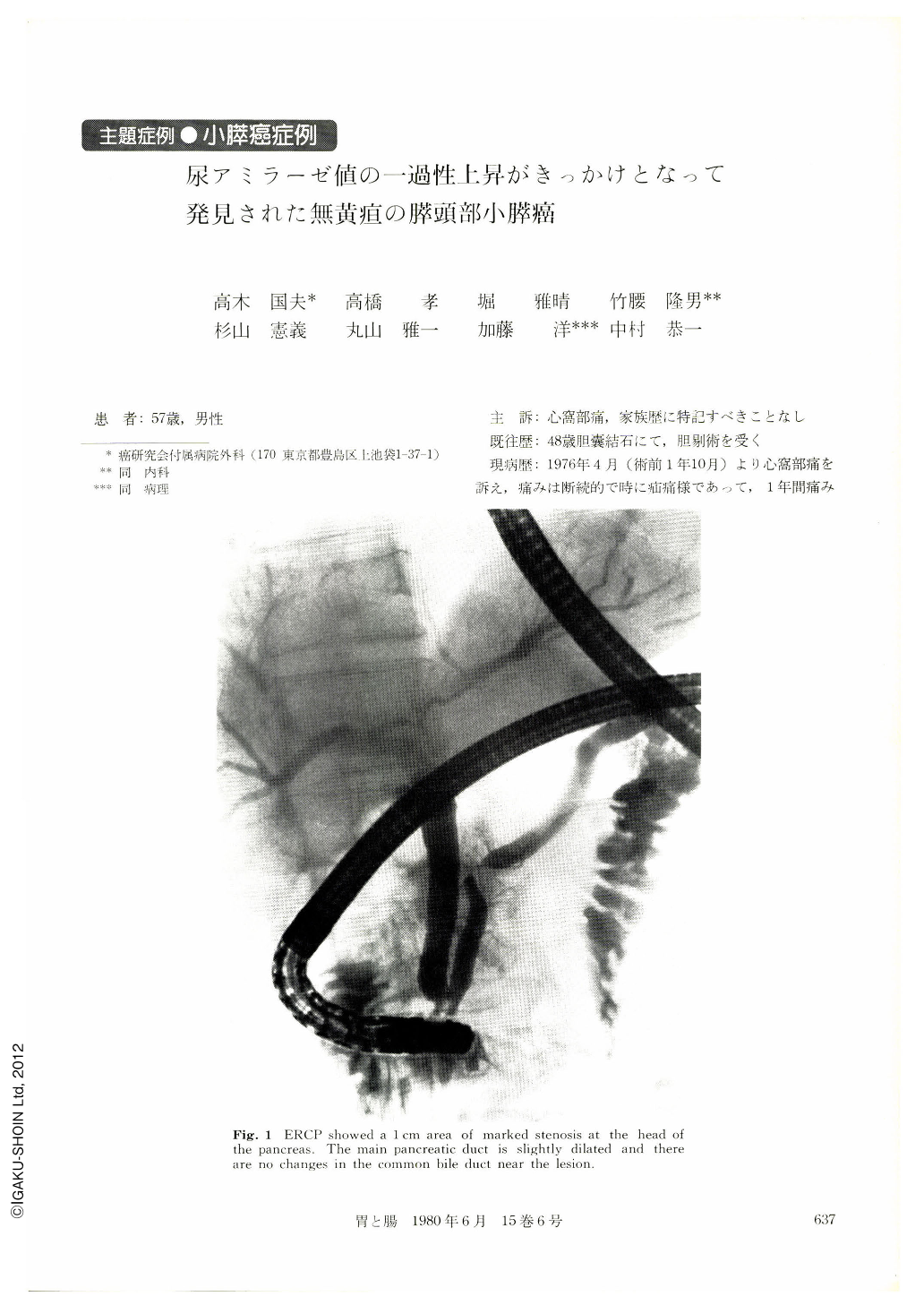
The patient is a 57 year-old male with carcinoma of the pancreas head. Nine years after cholecysteo tomy for gall stone he began to beel intermittent, colicky pain on the upper abdomen and in May, 1977, one year later after its onset, he visited the outpatient clinic of our hospital. There was no jaundice and laboratory examination revealed only a slight elevation on urinary amylase level, measuring 960 units (normal, less than 800 units) and no elevation of serum amylase, measuring 178 units. Scintigraphy of the pancreas showed low up-take of the entire pancreas. The patient visited the hospital again three months later and a weight loss of 5 kg was noted. Urinary amylase returned to normal (755 units). ERCP revealed a marked stenosis with length of 1cm at the pancreatic head. The proximal duct is dislocated to the common bile duct and looks rigitl and straightened (Fig. 1). Altliough the stenotic pancreatic cluct wall was relatively smooth, these findings led us to suspect the presence of a small malignant lesion. Pancreatic juice cytology performed twice during ERCP was negative for cancer cell.
The stenotic portion was further observed by inserting an encloscopically guided 2.3 mm diameter frontview fiberscope into the pancreatic duct. We used one of the recently developed peroral pancreatochclangioscope (Mother-and-baby fiberscope) and inserted the baby fiberscope into the pancreatic duct. Hemorrhage and slightly pale, granulated surface on the inner wall of stenotic pancreas duct at the papilla side were noted (Fig. 2). The lesion was thought to be pancreatic cancer invading the pancreatic duct. Ceiiac angicgraphy alone revealed no marked changes and simultaneous ceiiac angiography and ERCP discovered no marked changes in the intra-pancreatic arteries at the site of stenosis (Fig. 3).
Pancreaticoduodenectomy was performed on 15 th, February 1978. There was no ascites or adhesion in the abdominal cavity and no liver metastasis. On gross inspection, the resected specimen (Fig. 4) revealed a scarred, shallow depression on the pancreas capsule at the stenotic portion, but no mass was palpated. Near the stenosis, a slight induration was noted on the papilla side, but a macroscopic diagnosis of cancer was not possible. Histology (Fig. 5, 6, 7) revealed adenocarcinoma extending over an area measuring 2cm in diameter at the site of stenosis. The cancer tissue showing dendritic infiltration, was partly cystic, and limited in the pancreas. lntraductal spread of the adenocarcinoma along the inner wall of the duct may correspond to confirming the earlier pancreatoscopic findings. There was fibrosis in the pancreas head. The lymph nodes around the panareas were not involved. The patient's postoperative course was uneventful and he was discharged on the 25th postoperative day and two years later, he is well without recurrence.

Copyright © 1980, Igaku-Shoin Ltd. All rights reserved.


