Japanese
English
- 有料閲覧
- Abstract 文献概要
- 1ページ目 Look Inside
十二指腸から直腸におよぶ消化管を広範かつび慢性に侵したmultiple lymphomatous polyposisの稀有なる症例を経験したので報告する.
症 例
患 者:69歳 男性 公務員
主 訴:体重減少,全身倦怠感
家族歴:特記すべきことなし.子供3人の胃および大腸X線所見に異常なし.
既往歴:30歳,虫垂切除 現病歴:1977年2月より全身倦怠感および体重減少(1年間に6kg)に気づいた.6月14日,癌研内科受診.胃X線検査で十二指腸の異常所見を指摘され,精査のため6月30日入院した.
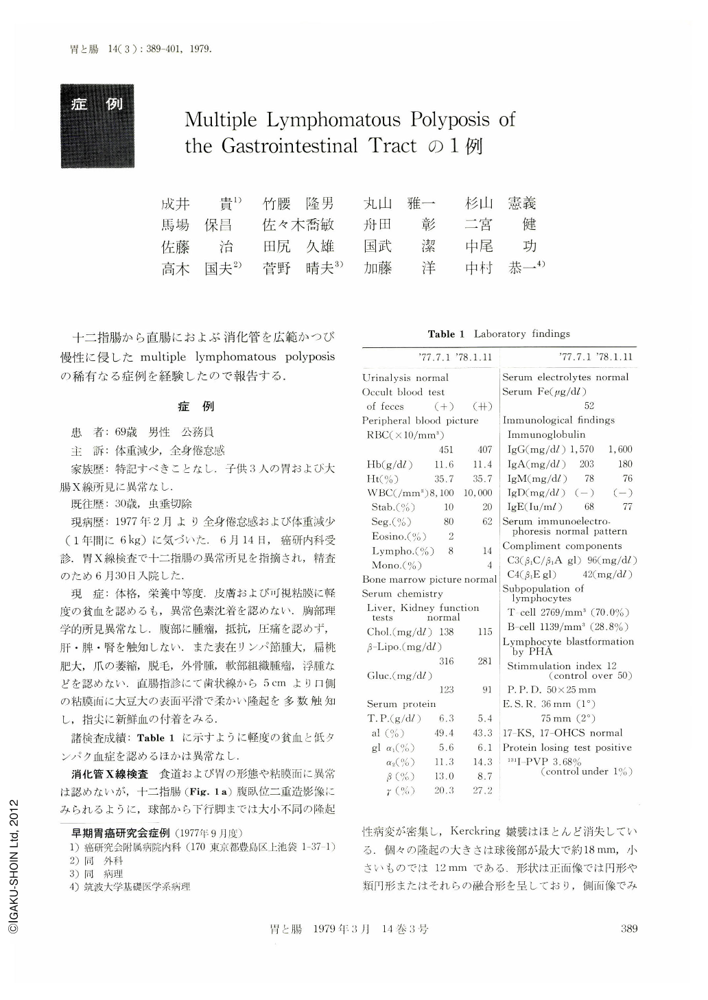
A 69-year-old male patient was admitted to the Department of Internal Medicine, Cancer Institute Hospital, Tokyo, with the chief complaint of weight loss and easy fatigability which had lasted for about six months. The physical examination showed unremarkable findings except for pallor of the skin and anemic conjunctiva of the palpebrae. Lymphoadenopathy, hepatosplenomegaly, palpable mass and pigmentation of the skin were not observed. On blood count hemoglobin was 11.6 g/dl, and white blood cell count was 8,100 with normal differentials. Serum total protein was 5.4 g/dl. No abnormality was seen in the findings of bone marrow puncture. Immunoelectrophoresis was normal. 131I-PVP test was positive with a value of 3.68% (control under 1%), suggestive of marked protein losing enteropathy. Subpopulations of T cell and B cell in the peripheral lymphocytes were normal. Blastformation of lympho-cytes induced by PHA was 12 (control over 50).
X-ray examination of the GI tract revealed innumerable polypoid lesions in the duodenum, jejunum, ileum and entire large bowel. Their form was mostly simulating those observed in submucosal tumor, and several larger ones were narrow based sessile lesions. Their sizes, measured in the largest diameters, were ranging from 2 to 20 mm in the duodenum, distal ileum and large bowel. These lesions were so densely distributed that there was no intervening mucosa between the lesions. In the proximal jejunum their distribution was rather rough and there was the larger area of the normal intervening mucosa with the Kerckring's folds. In the large bowel there was no deformation of the lumen with the normal haustrations. The mucosae of the esophagus and stomach appeared normal.
Biopsy materials were taken from the lesions of the duodenum by fiberduodenoscopy. Histologically these materials only revealed proliferation of small mature lymphocytes. Three lesions of the sigmoid colon were obtained by the technique of endoscopic polypectomy and the histological study was done. There was remarkable proliferation of small monomorphic mature lymphocytes in the mucosa and submucosa of the polypectomized materials with undistinguishable formation of the follicle germinal centers. The patient was discharged on 1 st of August, 1977 and has been placed under strict follow-up examination.
In this case the clinical and histological evidence available until present has led to the diagnosis of a benign nature which belongs to a disease entity called “multiple lymphomatous polyposis of the gastrointestinal tract” by Corner.

Copyright © 1979, Igaku-Shoin Ltd. All rights reserved.


