Japanese
English
- 有料閲覧
- Abstract 文献概要
- 1ページ目 Look Inside
- サイト内被引用 Cited by
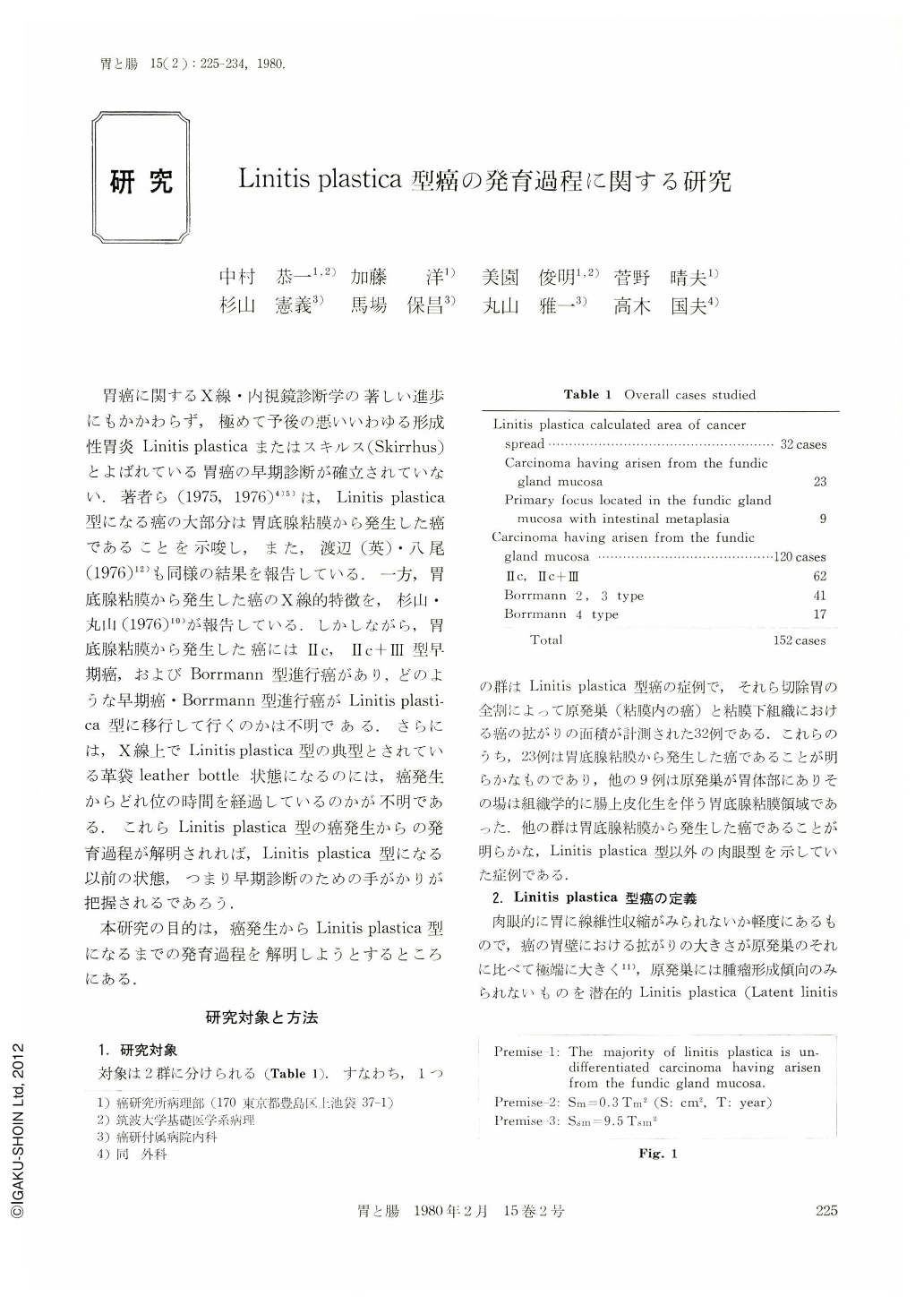
胃癌に関するX線・内視鏡診断学の著しい進歩にもかかわらず,極めて予後の悪いいわゆる形成性胃炎Linitis plasticaまたはスキルス(Skirrhus)とよばれている胃癌の早期診断が確立されていない.著者ら(1975,1976)4)5)は,Linitis plastica型になる癌の大部分は胃底腺粘膜から発生した癌であることを示唆し,また,渡辺(英)・八尾(1976)12)も同様の結果を報告している.一方,胃底腺粘膜から発生した癌のX線的特徴を,杉山・丸山(1976)10)が報告している.しかしながら,胃底腺粘膜から発生した癌にはHc,Hc+HI型早期癌,およびBorrmann型進行癌があり,どのような早期癌・Borrmann型進行癌がLinitis plastica型に移行して行くのかは不明である.さらには,X線上でLinitis plastica型の典型とされている革袋leather bottle状態になるのには,癌発生からどれ位の時間を経過しているのかが不明である.これらLinitis plastica型の癌発生からの発育過程が解明されれば,Linitis plastica型になる以前の状態,つまり早期診断のための手がかりが把握されるであろう.
本研究の目的は,癌発生からLinitis plastica型になるまでの発育過程を解明しようとするところにある.
The lapse of time and the morphological change from cancer-development to carcinoma of linitis plastica type have been studied in this paper.
Objects for this study consist of two groups (Table 1): one is 32 cases of the carcinoma of Iinitis plastica type whose primary focus was histologically clarified (Figs. 6~8) and whose areas of cancer spread to the mucosa and submucosa were estimated respectively, and another is 120 cases of carcinoma having arisen from the fundic gland mucosa (Figs. 3~5). Further, the primary focus of linitis plastica type means carcinoma limited to the mucosa. In 23 cases out of the 32 carcinomas of linitis plastica type, their primary foci were completely situated in the fundic gland mucosa without intestinal metaplasia, and primary foci of the remnant 9 cases in the fundic gland mucosa with intestinal metaplasia. All these resected stomachs were histologically examined by means of serially cutting method as shown in Figs. 3 and 6.
This study has started with the following three premises (Fig. 1): 1) Carcinoma arising from the fundic gland mucosa is histologically undifferentiated type (diffuse carcinoma), and the majority of carcinoma of linitis plastica type is that having arisen from the fundic gland mucosa2)6)); 2) The relation between the area of carcinoma spread in the mucosa (Sm cm2) and the lapse time of it from cancer-development (Tm year) is Sm=0.3 Tm25)6); 3) The relation between the area of carcinoma spread in the submucosa (Sm cm2) and the lapse time of it from the beginning of submucosal invasion (Tsm year) is Ssm=9.5 Tsm22)8).
Table 3 shows the mean values of the area and the lapse time in the 32 cases of carcinoma of linitis plastica type (the following abbreviates as L. P.). Sm and Ssm are values of actual measurement, and Tm and Tsm are calculated from these two values and premise-2 and premise-3. (Tm-Tsm) means the time when cancer infiltrated to the submucosa, and 0.3 (Tm-Tsm)2 means the area of the primary focus at that time.
In the 32 cases, 13 cases (Group A in Table 3) showed minus value in as shown in Table 2. Its finding is theoretically unreasonable, because it means that cancer develops in the submucosa. However, it demonstrates clearly that these 13 cases trated to the submucosa at the time when the area of the primary focus (Sm) measured less than 1.7cm in diameter. Residual 19 out of the 32 cases (Group B in Table 3) infiltrated to the submucosa at the time when the area of the primary focus measured 2.5 cm in diameter (0.3(Tm-Tsm)2). The average of those two values, 1.7 and 2.5cm, was less than 2cm.
The 13 cases belonging to Group A were macroscopically the stomach without constriction (Fig. 6) or with a slightly partial one. On the other hand. the 19 cases belonging to Group B showed macroscopically the stomach with moderate to marked constriction, named racliologically as “leather bottle”.
The size of carcinoma spread in the mucosa is generally considered as a scale showing the lapse of time from cancer-development. Table 4 shows the relationship between the average diameter of carcinoma spread in the mucosa and macroscopic type of the objects. The average diameter of 62 carcinomas of Type Ⅱc and Type Ⅱc+Ⅲ (2.48 cm) in the early phase is smaller than that of 58 carcinomas of Borrmann type (3.77 cm) and that of 32 L. P. (3.68 cm) in advanced phase. However, the average diameter of the carcinoma of the Borrmann type is almost similar to that of the L. P. The following conclusions are drawn from the data obtained: The lapse time of carcinoma in early phase is generally shorter than that in advanced phase, and it may be interpreted that the majority of L. P. progresses immediately from early carcinoma without passing through the Borrmann type, because the size of carcinoma of the Borrmann type and L. P. spreading in the mucosa is almost same. If L. P. progresses via the Borrmann type, the size should be different.
Out of the objects, 52 carcinomas whose intramucosal spread measures less than 2 cm in diameter were macroscopically examined on the convergency of the mucosal fold in its primary focus (Table 5). About 70% of 31 carcinomas in early phase and 12 carcinomas of Borrmann type had convergency of the mucosa] fold, however, only one case had it in 9 cases of L. P. Therefore, it may be suggested that intramucosal carcinoma progresses to L. P. on the occasion of which it infiltrated to the submucosa before cancerous ulceration, because convergency of the mucosal fold is attributable to ulcerative fibrosis.
A hypothesis on growing process to carcinoma of linitis plastica type from cancer-development may be led from the data mentioned above as follows: Carcinoma developed in the fundic gland mucosa infiltrates to the submucosa antecedently to cancerous ulceration. Area of the carcinoma at the time of submucosal infiltration measures about 2cm or less than 2cm in diameter (Fig. 9). After that, ulceration may occur in the carcinoma, because undifferentiated carcinoma (diffuse carcinoma) has a striking tendency to easily ulcerate with increasing size of the intramucosal carcinoma8)10). The carcinoma that measures approximately 2cm in diameter of intramucosal extent, that spreads widely in the submucosa, and that does not yet show constriction of the stomach, may have a lapse of about 3 years after cancer-development (Latent linitis plastica). Areas of the carcinoma increase in the mucosa and submucosa with the lapse of time, independently of each other. Whereas, fibrosis accompaning with cancer infiltration increases gradually in the submucosa and proper muscle, and constriction of the stomach occurs (Typical linitis plastica, named radiologically as “leather bottle”).
According to this hypothesis, early detection of carcinoma of linitis plastica type is to diagnose carcinoma of Type Ⅱc without converge-ncy of the mucosal fold, the location of which is histologically the fundic gland mucosa without intestinal metaplasia or with one of slight degree, and macroscopically the gastric body with many mucosal folds.

Copyright © 1980, Igaku-Shoin Ltd. All rights reserved.


