Japanese
English
- 有料閲覧
- Abstract 文献概要
- 1ページ目 Look Inside
- サイト内被引用 Cited by
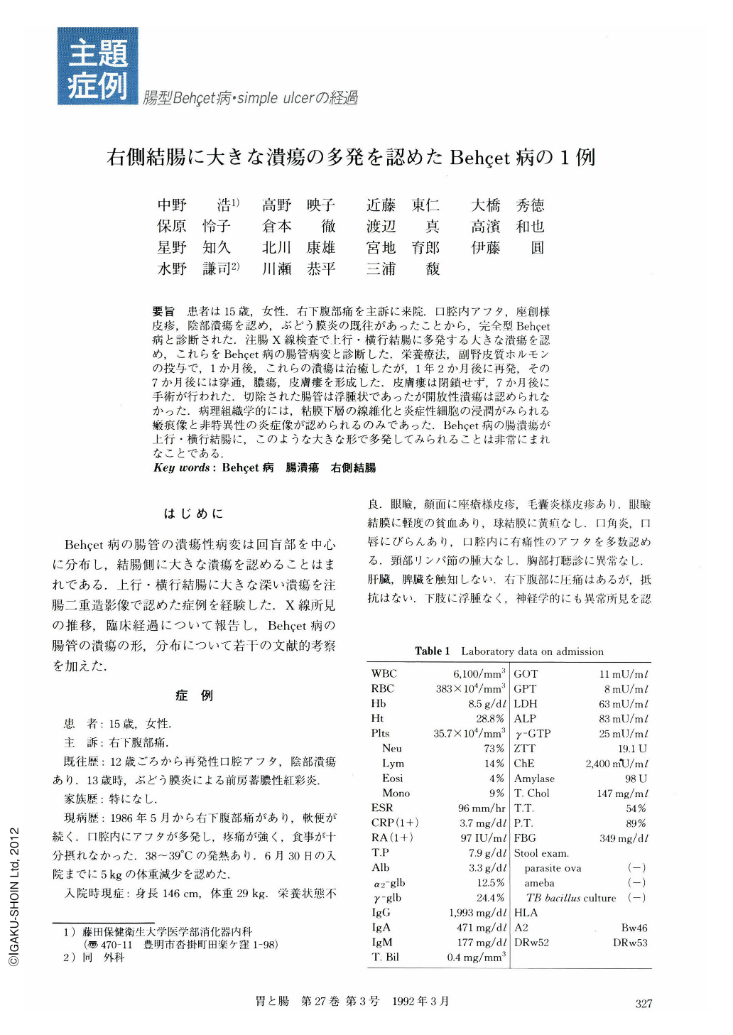
要旨 患者は15歳,女性.右下腹部痛を主訴に来院.口腔内アフタ,座創様皮疹,陰部潰瘍を認め,ぶどう膜炎の既往があったことから,完全型Behçet病と診断された.注腸X線検査で上行・横行結腸に多発する大きな潰瘍を認め,これらをBehçet病の腸管病変と診断した.栄養療法,副腎皮質ホルモンの投与で,1か月後,これらの潰瘍は治癒したが,1年2か月後に再発,その7か月後には穿通,膿瘍,皮膚瘻を形成した.皮膚瘻は閉鎖せず,7か月後に手術が行われた.切除された腸管は浮腫状であったが開放性潰瘍は認められなかった.病理組織学的には,粘膜下層の線維化と炎症性細胞の浸潤がみられる瘢痕像と非特異性の炎症像が認められるのみであった.Behçet病の腸潰瘍が上行・横行結腸に,このような大きな形で多発してみられることは非常にまれなことである.
A 15-year-old female with the chief complaint of right lower abdominal pain visited our clinic. She had recurrent aphthoid ulcers in her oral cavity and acneiform skin eruptions, genital ulcer and an episode of uvenitis. She was diagnosed as complete type of Behçet's disease.
Radiological examination of the colon revealed multiple, large ulcers in the ascending colon and transverse colon and a small ulcer in the terminal ileum. Those ulcerative lesions of the colon and ileum were diagnosed as the intestinal lesions of Behçet's disease.
After nutritional therapy and administration of prednisolone, the ulcerations disappeared within two months. One year and two months later, those intestinal ulcers reccurred again. Skin fistula was found through abdominal abscess due to penetration of the ulcer in the ascending colon. This fistula was not closed for seven months and surgical operation with right hemicolectomy was performed.
Macroscopic findings of the resected specimen showed edematous and thikened wall and ulcer scars on the mucosal surface. Histological examination revealed a thin mucosal layer and ulcer scar with fibrosis and infiltration of inflammatory cells to the submucosal layer.
It is very uncommon to find those large, discrete ulcers in the right colon in Behçet's disease.
Copyright © 1992, Igaku-Shoin Ltd. All rights reserved.