Japanese
English
- 有料閲覧
- Abstract 文献概要
- 1ページ目 Look Inside
- サイト内被引用 Cited by
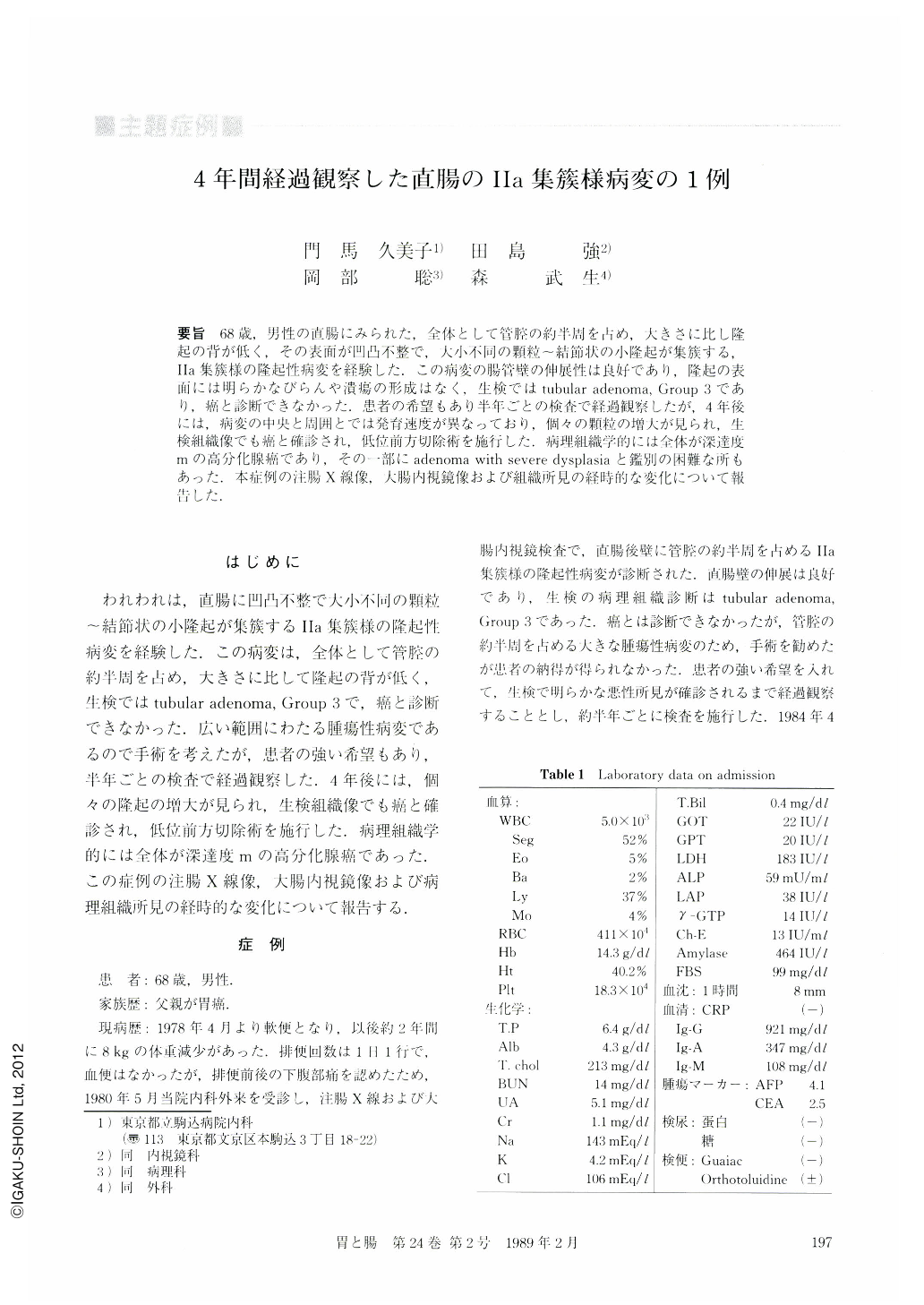
要旨 68歳,男性の直腸にみられた,全体として管腔の約半周を占め,大きさに比し隆起の背が低く,その表面が凹凸不整で,大小不同の顆粒~結節状の小隆起が集簇する,Ⅱa集簇様の隆起性病変を経験した.この病変の腸管壁の伸展性は良好であり,隆起の表面には明らかなびらんや潰瘍の形成はなく,生検ではtubular adenoma,Group 3であり,癌と診断できなかった.患者の希望もあり半年ごとの検査で経過観察したが,4年後には,病変の中央と周囲とでは発育速度が異なっており,個々の顆粒の増大が見られ,生検組織像でも癌と確診され,低位前方切除術を施行した.病理組織学的には全体が深達度mの高分化腺癌であり,その一部にadenoma with severe dysplasiaと鑑別の困難な所もあった.本症例の注腸X線像,大腸内視鏡像および組織所見の経時的な変化について報告した.
A 68-year-old male patient with a Ⅱa-aggregated-like lesion in the rectum was followed-up for four years. Endoscopical and barium enema studies demonstrated gradual increase in both width and height, with the width increasing from 6 to 7 cm during the follow-up period. The central nodular lesion changed most markedly compared with the surrounding granular lesions. Four years later adenocarcinoma was diagnosed in the biopsy specimen taken from the central nodular lesion. Surgical material, 5×3.5 cm in size, had large Ⅱa-aggregated-like lesion. The center of the lesion differed from the rest of the area in that the surface lacked granular appearance.
Histological examination revealed adenocarcinoma in part of the lesion and various degrees of structural and cellular atypism in the rest of the area of the lesion. Cancer was confined only to the mucosa.

Copyright © 1989, Igaku-Shoin Ltd. All rights reserved.


