Japanese
English
- 有料閲覧
- Abstract 文献概要
- 1ページ目 Look Inside
- サイト内被引用 Cited by
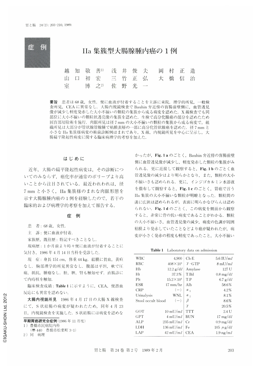
要旨 患者は68歳,女性.便に血液が付着することを主訴に来院.理学的所見,一般検査所見,CEAに異常なし.大腸内視鏡検査でBauhin弁近傍の盲腸前壁側に,血管透見像が減少し軽度発赤した大小不揃いの顆粒の集簇から成る病変を認めた.X線検査でも同部位に大小不揃いの顆粒状透亮像の集簇を認めた.生検で高分化腺癌の部分を認めたため回盲部切除術を施行.肉眼所見は径7mmの大小不揃いの顆粒の集簇から成る病変で,組織所見は大部分が管状腺管腺腫で粘膜表層の一部に高分化管状腺癌を認めた.径7mmと小さなⅡa集簇様病変の術前診断例はまれであり,X線,内視鏡所見を中心に呈示し,大腸扁平隆起性病変に関する臨床病理学的考察を加えた.
A 68-year-old woman who had occasionally noticed fresh blood on stool and visited our hospital. Laboratory examination showed no abnormalities (Table 1). Colonoscopy showed a small flat elevated lesion composed of multiple conglomerated small nodules on the anterior wall of the cecum (Fig. 1). Barium enema also showed the similar lesion corresponding to the colonoscopic findings (Fig. 2). Biopsy specimens were interpreted as focal adenocarcinoma in tubular adenoma (Fig. 3). Ileo-cecal colectomy was performed. Macroscopic diagnosis was Ⅱa conglomerated type cancer of the large intestine, measuring 7×7 mm in diameter (Fig. 4). Histological diagnosis was focally well-differentiated adenocarcinoma in tubular adenoma limited to the mucosa (Fig. 5).
Ⅱa conglomerated type colon cancer is extremely rare and difficult to diagnose. This case is considered to be valuable in suggesting histogenesis of the cancer of the large intestine.
Copyright © 1989, Igaku-Shoin Ltd. All rights reserved.