Japanese
English
- 有料閲覧
- Abstract 文献概要
- 1ページ目 Look Inside
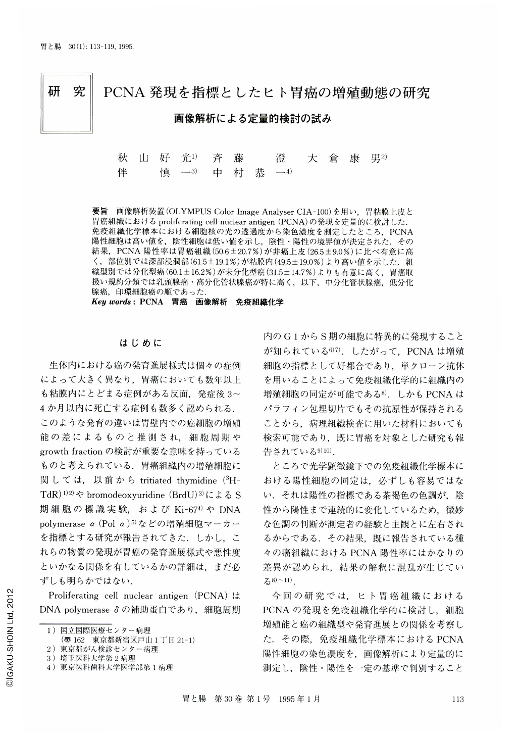
要旨 画像解析装置(OLYMPUS Color Image Analyser CIA-100)を用い,胃粘膜上皮と胃癌組織におけるproliferating cell nuclear antigen(PCNA)の発現を定量的に検討した.免疫組織化学標本における細胞核の光の透過度から染色濃度を測定したところ,PCNA陽性細胞は高い値を,陰性細胞は低い値を示し,陰性・陽性の境界値が決定された.その結果,PCNA陽性率は胃癌組織(50.6±20.7%)が非癌上皮(26.5±9.0%)に比べ有意に高く,部位別では深部浸潤部(61.5±19.1%)が粘膜内(49.5±19.0%)より高い値を示した.組織型別では分化型癌(60.1±16.2%)が未分化型癌(31.5±14.7%)よりも有意に高く,胃癌取扱い規約分類では乳頭腺癌・高分化管状腺癌が特に高く,以下,中分化管状腺癌,低分化腺癌,印環細胞癌の順であった.
Expression of proliferating cell nuclear antigen (PCNA) in 42 cases of gastric carcinoma was studied quantitatively using a color image analysis system (OLYMPUS CIA-100). Non-cancerous gastric mucosa and gastric cancer tissues were stained immunohistochemically with a monoclonal antibody against PCNA. Subsequently, the intensity of the nuclear color was analyzed by the degree of light permeability in CIA - 100 to determine positivity or negativity. The mean value of the positive cell rate (% PCNA) of the carcinomas was significantly higher (50.6±20.7%, p<0.01) than that of the non-cancerous epithelia (26.5%±9.0%). The cancer cells infiltrating deeper layers of the gastric wall exhibited a higher %PCNA (61.5%±19.1%, p<0.01) than those located in the lamina propria of the mucosa (49.5%±19.0%). The differentiated carcinomas showed a significantly higher %PCNA (60.1%±16.2%, p<0.01) than the undifferentiated carcinomas (31.5%±14.7%). According to the histological classif ication of The General Rules for the Gastric Cancer Study (Japanese Research Society for Gastric Cancer), pap+tub1 showed the highest %PCNA, followed by tub 2, por and sig, successively.

Copyright © 1995, Igaku-Shoin Ltd. All rights reserved.


