Japanese
English
- 有料閲覧
- Abstract 文献概要
- 1ページ目 Look Inside
- 参考文献 Reference
- サイト内被引用 Cited by
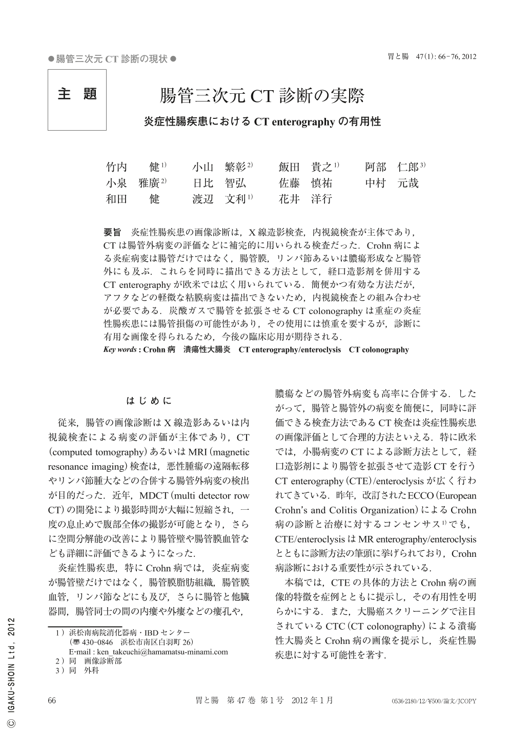
要旨 炎症性腸疾患の画像診断は,X線造影検査,内視鏡検査が主体であり,CTは腸管外病変の評価などに補完的に用いられる検査だった.Crohn病による炎症病変は腸管だけではなく,腸管膜,リンパ節あるいは膿瘍形成など腸管外にも及ぶ.これらを同時に描出できる方法として,経口造影剤を併用するCT enterographyが欧米では広く用いられている.簡便かつ有効な方法だが,アフタなどの軽微な粘膜病変は描出できないため,内視鏡検査との組み合わせが必要である.炭酸ガスで腸管を拡張させるCT colonographyは重症の炎症性腸疾患には腸管損傷の可能性があり,その使用には慎重を要するが,診断に有用な画像を得られるため,今後の臨床応用が期待される.
Radiographic and endoscopic examinations are often used for the diagnosis and evaluation of IBD(inflammatory bowel diseases), while CT(computed tomography)is considered a complementary investigation for extra-intestinal lesions in CD(Crohn's disease), such as fistulae and abscesses. Further, in Europe and the United States, CTE(CT enterography), which is a contrast enhanced CT scan with oral neutral contrast agents for dilatation of the intestine, is an attractive new technique for the diagnosis of CD. CTE can provide graphical images from both, the bowel wall and extra-intestinal lesions of CD, and has been associated with a high level of tolerance among patients. However, endoscopic examination is necessary for the detection of aphthous ulcers as these can not be detected by CTE. Additionally, while CTC(CT colonoscopy)can produce diagnostic images of IBD lesions, some perforations in IBD patients associated with the application of CTC have been reported. Accordingly, although, further modification of this technique appears desirable for IBD patients, it is hoped that CTC will become routine in clinical settings.

Copyright © 2012, Igaku-Shoin Ltd. All rights reserved.


