Japanese
English
- 有料閲覧
- Abstract 文献概要
- 1ページ目 Look Inside
- サイト内被引用 Cited by
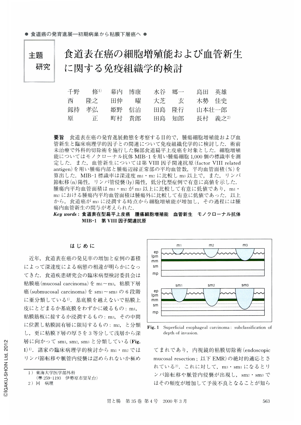
要旨 食道表在癌の発育進展動態を考察する目的で,腫瘍細胞増殖能および血管新生と臨床病理学的因子との関連について免疫組織化学的に検討した.術前未治療で外科的切除術を施行した胸部食道扁平上皮癌を対象とした.細胞増殖能についてはモノクローナル抗体MIB-1を用い腫瘍細胞1,000個の標識率を測定した.また,血管新生については第VIII因子関連抗原(factor VIII related antigen)を用い腫瘍内部と腫瘍辺縁正常部の平均血管数,平均血管面積(%)を算出した.MIB-1標識率は深達度m1・m2に比較しm3以上で,また,リンパ節転移(n)陽性,リンパ管侵襲(ly)陽性,低分化型症例で有意に高値を示した.腫瘍内平均血管面積はm1・m2がm3以上に比較して有意に低値であり,m1・m2における腫瘍内平均血管面積は腫瘍外に比較して有意に低値であった.以上から,食道癌がm3に浸潤する時点から細胞増殖能が増加し,その過程には腫瘍内血管新生の関与が考えられた.
The correlation of clinicopathologcal characteristics with proliferative activity and angiogenesis was investigated immunohistochemically to assess the growth of superficial esophageal carcinoma. The cases of squamous cell carcinoma in the thoracic esophagus which underwent surgical resecion without preoperative treatment were studied. The labeling index (LI) to evaluate the proliferative activity of MIB-1 was calculated as the number of positive nuclei per 1,000 cancer cells counted. Factor VIII-related antigen was used to measure the mean number of blood vessels and blood-vessel surface area (%) inside the tumor (intratumor) and in the normal tissue around the tumor (extratumor) . The LI of MIB-1 of depth m3 carcinoma was significantly higher than that of m1or m2 carcinoma. The LI of poorly differentiated carcinoma was significantly higher than that of well or moderately differentiated carcinoma. The LI of cases of lymph node metastasis or lymphatic invasion was significantly higher than in cases without it. The mean intratumor blood-vessel surface area (%) was significantly smaller in m1 or m2 lesions than in lesions with greater depths. These results suggested that the growth of esophageal carcinoma may accelerate after the invasion of the muscularis mucosa (depth m3) with the increase of proliferative activity. Intratumor angiogenesis may contribute to the process by which superficial esophageal carcinoma grows from m1/m2 to m3.

Copyright © 2000, Igaku-Shoin Ltd. All rights reserved.


