Japanese
English
- 有料閲覧
- Abstract 文献概要
- 1ページ目 Look Inside
- 参考文献 Reference
- サイト内被引用 Cited by
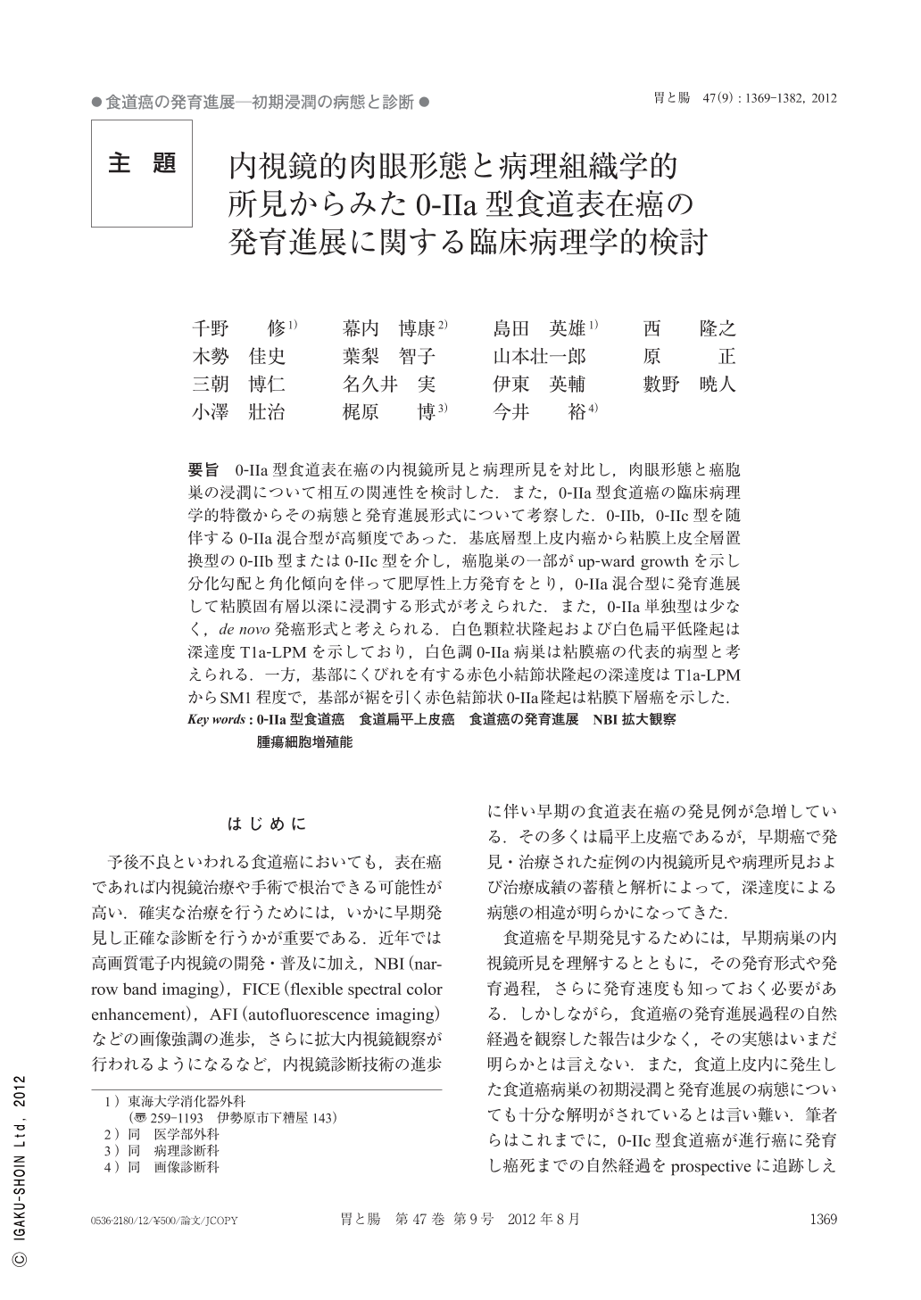
要旨 0-IIa型食道表在癌の内視鏡所見と病理所見を対比し,肉眼形態と癌胞巣の浸潤について相互の関連性を検討した.また,0-IIa型食道癌の臨床病理学的特徴からその病態と発育進展形式について考察した.0-IIb,0-IIc型を随伴する0-IIa混合型が高頻度であった.基底層型上皮内癌から粘膜上皮全層置換型の0-IIb型または0-IIc型を介し,癌胞巣の一部がup-ward growthを示し分化勾配と角化傾向を伴って肥厚性上方発育をとり,0-IIa混合型に発育進展して粘膜固有層以深に浸潤する形式が考えられた.また,0-IIa単独型は少なく,de novo発癌形式と考えられる.白色顆粒状隆起および白色扁平低隆起は深達度T1a-LPMを示しており,白色調0-IIa病巣は粘膜癌の代表的病型と考えられる.一方,基部にくびれを有する赤色小結節状隆起の深達度はT1a-LPMからSM1程度で,基部が裾を引く赤色結節状0-IIa隆起は粘膜下層癌を示した.
We analyzed the relationship between the macroscopic view and the invasive level of the cancer by endoscopic findings of the lesion and its pathological findings on type 0-IIa superficial esophageal carcinoma. Moreover we discussed the status and way of development of the lesion according to the clinicopathological features of type 0-IIa cancer. Type 0-IIa cancer was frequently accompanied by type 0-IIb or 0-IIc. A part of the cancer lesion seems to have an elevating hypertrophic and keratinizing tendency showing upward growth, which is identified as 0-IIa, and to a similar degree invades deeper beneath the layer of the lamina propria mucosae. The pure type of the 0-IIa lesion is rarely encountered, and it seems to appear as a de novo cancer. When the lesions elevated portion is whitish and granular or presents a whitish, low, flat elevation, the depth of invasion is M2. a whitish lesion of type 0-IIa shows typically a invasion limited to the mucosa. On the other hand, further invasion as in M2 to SM1 is suggested for the reddish small granular lesion constrictor at the base, and SM is suggested for 0-IIa type of red granulation with smooth elevation. The nodal part of the red protrusion seems to invade deeper to the submucosal layer with the gentle sloping.

Copyright © 2012, Igaku-Shoin Ltd. All rights reserved.


