Japanese
English
- 有料閲覧
- Abstract 文献概要
- 1ページ目 Look Inside
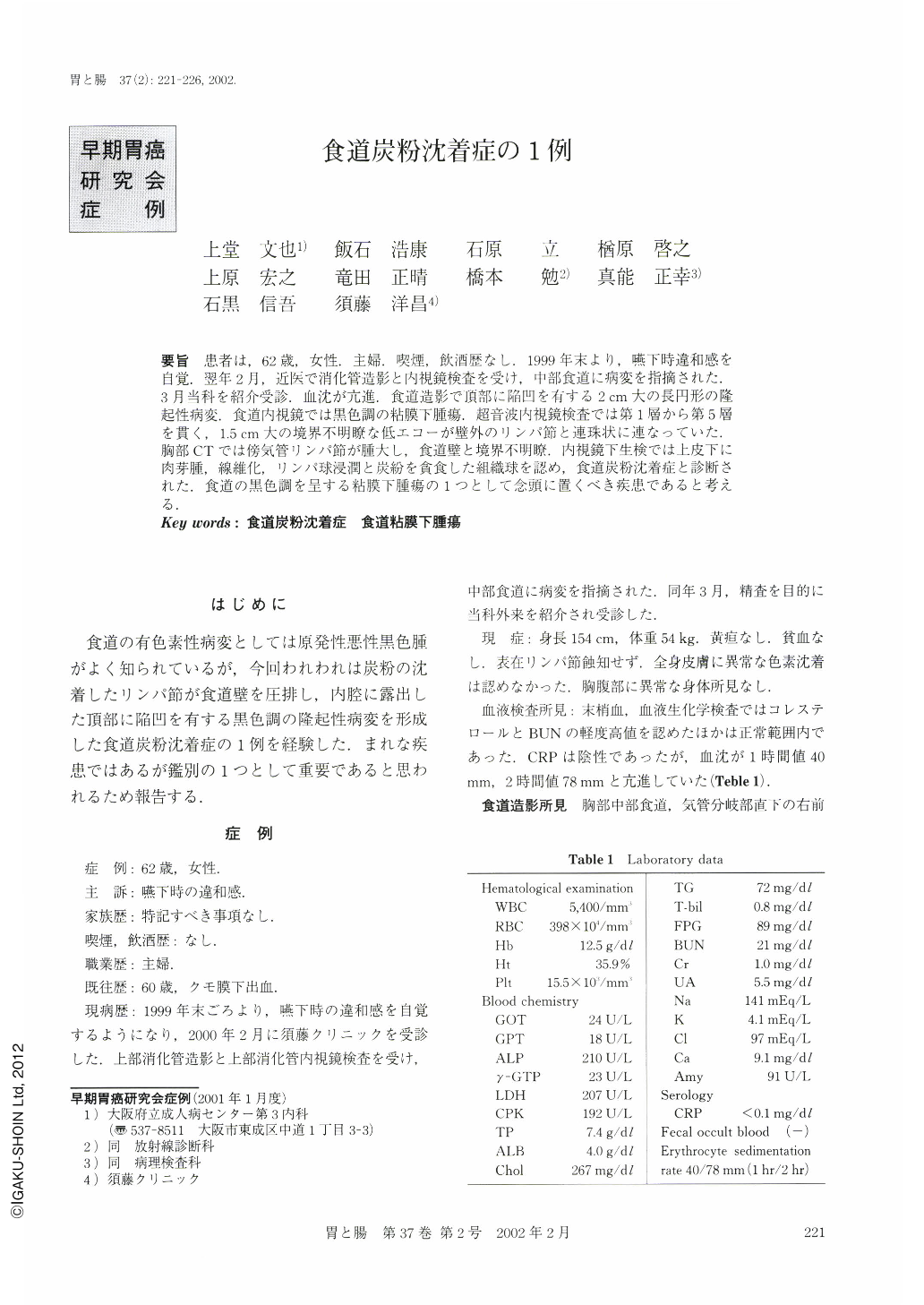
要旨 患者は,62歳,女性.主婦.喫煙,飲酒歴なし.1999年末より,嚥下時違和感を自覚.翌年2月,近医で消化管造影と内視鏡検査を受け,中部食道に病変を指摘された.3月当科を紹介受診.血沈が亢進.食道造影で頂部に陥凹を有する2cm大の長円形の隆起性病変.食道内視鏡では黒色調の粘膜下腫瘍.超音波内視鏡検査では第1層から第5層を貫く,1.5cm大の境界不明瞭な低エコーが壁外のリンパ節と連珠状に連なっていた.胸部CTでは傍気管リンパ節が腫大し,食道壁と境界不明瞭.内視鏡下生検では上皮下に肉芽腫,線維化,リンパ球浸潤と炭紛を貪食した組織球を認め,食道炭粉沈着症と診断された.食道の黒色調を呈する粘膜下腫瘍の1つとして念頭に置くべき疾患であると考える.
A 62 year-old-woman presented to her physician because of swallowing discomfort that had continued during a period of two months. She had no habit of drinking or smoking. Barium study and Esophago-gastroduodenoscopy disclosed an elevated lesion in the middle thoracic esophagus and she was referred to our hospital. Her erythrocyte sedimentation rate was elevated. Esophagography showed an oval elevated lesion with a central sharp depression. Esophagoscopy showed a black submucosal tumor and it was suspected to be an esophageal malignant melanoma. Endoscopic ultrasound demonstrated a low echoic area that extended to the extraluminal lymph nodes and penetrated the esophageal wall. Endoscopic biopsy specimen consisted of subepithelial granulomas, fibrosis, mononuclear inflammatory cell infiltration and it contained macrophages that phagocytosed coal pigments. It was diagnosed as esophageal anthracosis, which is rare but which is also important for differential diagnosis from esophageal submucosal tumors.

Copyright © 2002, Igaku-Shoin Ltd. All rights reserved.


