Japanese
English
- 有料閲覧
- Abstract 文献概要
- 1ページ目 Look Inside
- 参考文献 Reference
- サイト内被引用 Cited by
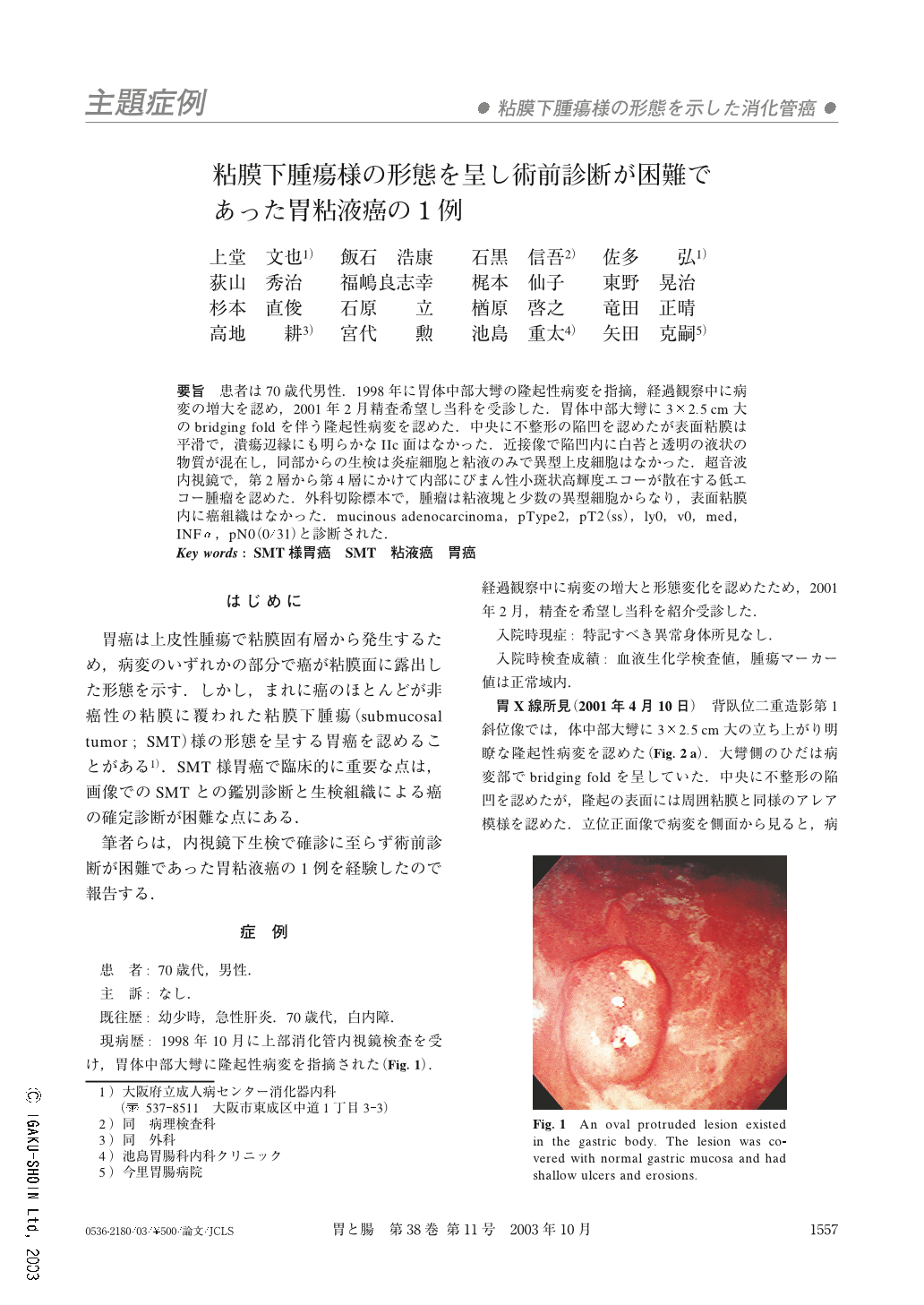
要旨 患者は70歳代男性.1998年に胃体中部大彎の隆起性病変を指摘,経過観察中に病変の増大を認め,2001年2月精査希望し当科を受診した.胃体中部大彎に3×2.5cm大のbridging foldを伴う隆起性病変を認めた.中央に不整形の陥凹を認めたが表面粘膜は平滑で,潰瘍辺縁にも明らかなIIc面はなかった.近接像で陥凹内に白苔と透明の液状の物質が混在し,同部からの生検は炎症細胞と粘液のみで異型上皮細胞はなかった.超音波内視鏡で,第2層から第4層にかけて内部にびまん性小斑状高輝度エコーが散在する低エコー腫瘤を認めた.外科切除標本で,腫瘤は粘液塊と少数の異型細胞からなり,表面粘膜内に癌組織はなかった.mucinous adenocarcinoma,pType2,pT2(ss),ly0,v0,med,INFα,pN0(0/31)と診断された.
A 74-year-old man had a stomachache and underwent esophago-gastro-duodenoscopy (EGD) 28 months ago. A protruded tumor, 2 cm in size, with small shallow ulcers on its surface was found in the gastric body. The lesion increased in size and the ulcers enlarged during the observation period, so he was referred to our hospital for further examination and treatment. Barium meal study showed a broad based protruded tumor with an irregular ulcer on its surface. EGD revealed that the tumor was covered with normal gastric mucosa and had an irregular ulceration. There was no “IIc” component, which indicates intramucosal carcinoma, on the surface mucosa. Biopsy specimens that were taken from the ulceration contained no malignant cells but did contain mucin and inflammatory cells. We diagnosed it as a malignant submucosal tumor (SMT) and the patient underwent an operation. The tumor was 2.5 cm in size. It had invaded to the subserosa and was covered with normal gastric mucosa. Histologically, it consisted of rich mucin and a few cancer cells and it was diagnosed as mucinous adenocarcinoma of the stomach. There was no cancer tissue in the surface mucosa, which made it difficult to make a diagnosis by biopsy. Mucinous adenocarcinoma of the stomach sometimes appears as an SMT-like gastric carcinoma. It is important to make a diagnosis according to its endoscopic appearance and mucin in the biopsy specimen. Biopsy may be negative because of its normal surface mucosa and rich mucous component.

Copyright © 2003, Igaku-Shoin Ltd. All rights reserved.


