Japanese
English
- 有料閲覧
- Abstract 文献概要
- 1ページ目 Look Inside
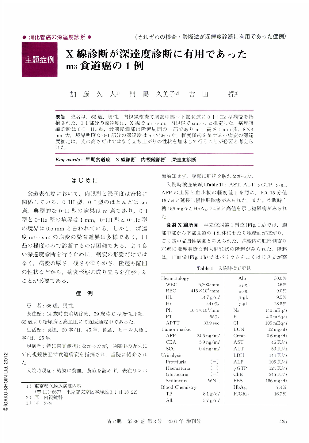
要旨 患者は,66歳,男性.内視鏡検査で胸部中部~下部食道に0-Ⅰ+Ⅱc型病変を指摘された.0-Ⅰ部分の深達度は,X線でm3~sm1,内視鏡でsm1~2と推定した.病理組織診断は0-Ⅰ+Ⅱc型,最深浸潤部は隆起周囲の一部でありm3.高さ1mm強,8×4mm大,境界明瞭な0-Ⅰ部分の深達度はm2であった.軽度隆起を呈する小病変の深達度推定は,丈の高さだけではなく立ち上がりの性状を加味して行うことが必要と考えられた.
We describe the usefulness of esophagography for estimating the depth of cancer invasion of early esophageal carcinoma in a 66-year-old man. A superficial and protruding lesion accompanied by a slight depression (type 0-Ⅰ+Ⅱc) from the middle to the lower thoracic esophagus was detected endoscopically. Endoscopic diagnosis was carcinoma massively invading the submucosa. On the other hand, esophagography showed a well-defined contour and lower height of the protrusion than estimated by endoscopy and no deformity of the esophageal margin. Radiologic diagnosis was carcinoma with invasion confined to the muscularis mucosae (m3) below the protrusion. The resected specimen showed a 0.8×0.4 cm-sized protruding lesion accompanied by a 5.5×3.5 cm-sized slightly depressed lesion. Histological examination revealed that cancer invasion below the protrusion remained in the lamina propria mucosae (m2), but the deepest point of invasion reached into the muscularis mucosae in a small area in the slightly depressed lesion. In conclusion, for accurate estimation of cancer invasion below protruding lesions, it is necessary to observe the contour of elevation in addition to the height of the protrusion and to take note of whether or not there is deformity of the esophageal margin.

Copyright © 2001, Igaku-Shoin Ltd. All rights reserved.


