Japanese
English
- 有料閲覧
PPV購入案内
1,320円 (1,200円+税10%) こちらは、1311ページから1320ページの文献です。購入には医書.jp本会員登録が必要です。
会員登録完了後に改めて上記「購入サイトへ」を選択してください。
または「購入サイトへ」選択後に「購入ログイン」、「新規会員登録」の順に進んでください。
会員登録について詳しくはをご確認ください。
- Abstract 文献概要
- 1ページ目 Look Inside
- 参考文献 Reference
- サイト内被引用 Cited by
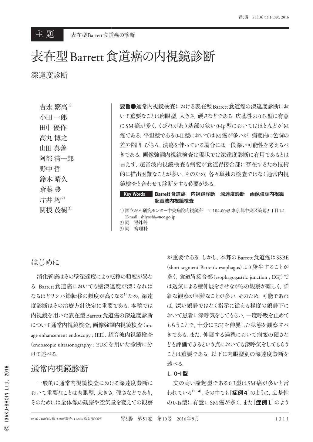
要旨●通常内視鏡検査における表在型Barrett食道癌の深達度診断において重要なことは肉眼型,大きさ,硬さなどである.広基性の0-Is型に有意にSM癌が多く,くびれがあり基部の狭い0-Ip型においてはほとんどがM癌である.平坦型である0-II型においてはM癌が多いが,病変内に色調の差や陥凹,びらん,潰瘍を伴っている場合には一段深い可能性を考えるべきである.画像強調内視鏡検査は現状では深達度診断に有用であるとは言えず,超音波内視鏡検査も病変が食道胃接合部に存在するため技術的に描出困難なことが多い.そのため,各々単独の検査ではなく通常内視鏡検査と合わせて診断をする必要がある.
During endoscopic diagnosis by conventional endoscopy, endoscopic morphology, size, and stiffness of lesions are important to estimate the depth of invasion. Sessile 0-I lesions tend to invade the submucosal layer, and pedunculated 0-I lesions are usually confined to the mucosa. Although flat lesions, such as 0-IIa or 0-IIc, are mainly mucosal cancers, we should consider that lesions may invade more deeply if they have differentiation of color, deeper depression, erosion, and ulceration. Thus far, there is no strong evidence that imaged enhanced endoscopy is useful for depth diagnosis. It can be difficult for EUS(endoscopic ultrasound)to reveal the submucosal invasion precisely because of their location. Therefore, we should diagnose using both EUS and conventional endoscopy.
Copyright © 2016, Igaku-Shoin Ltd. All rights reserved.