Japanese
English
- 有料閲覧
PPV購入案内
1,320円 (1,200円+税10%) こちらは、465ページから470ページの文献です。購入には医書.jp本会員登録が必要です。
会員登録完了後に改めて上記「購入サイトへ」を選択してください。
または「購入サイトへ」選択後に「購入ログイン」、「新規会員登録」の順に進んでください。
会員登録について詳しくはをご確認ください。
- Abstract 文献概要
- 1ページ目 Look Inside
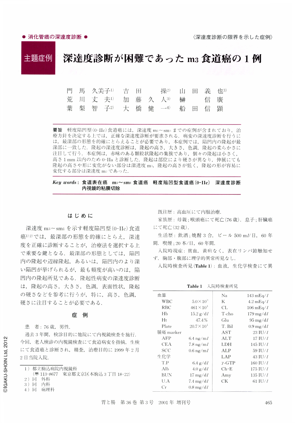
要旨 軽度陥凹型(0-Ⅱc)食道癌には,深達度m1~sm3までの症例が含まれており,治療方針を決定する上では,正確な深達度診断が要求される.病変の深達度診断を行うには,最深部の形態を的確にとらえることが必要であり,本症例では,陥凹内の隆起が最深部に一致した.隆起の深達度診断は,隆起の高さ,大きさ,色調,隆起の柔らかさに注目して行う.本症例は,赤味のある顆粒状隆起の集簇であり,個々の隆起は小さく,高さ1mm以内のため0-Ⅱaと診断した.隆起は部位により硬さが異なり,伸展にても隆起の高さや形に変化がない部分は深達度m3,隆起の高さが低く,隆起の形が容易に変化する部分は深達度m2であった.
A 76-year-old man was admitted to our hospital because of a superficial esophageal cancer which was detected during an annual examination of the upper gastrointestinal tract. An endoscopic study revealed a slightly depressed mucosal lesion with reddening, which occupied a quarter of the circumference at 30 cm from the incisor. Several granular changes in the lesion was also identified. It was classified as a type 0-Ⅱa+Ⅱc cancer with slight invasion into the submucosa (sm1), because granular changes showed no deformity during stretching of the esophageal wall at the time of the endoscopy. An esophagogram revealed a slight depression in the middle third of the esophagus, 3 cm long and extending over size and one fourth of the circumference. Partial irregularity of the lesion with deeper depression in the center of the irregular part suggested slight cancer invasion into the submucosa (sm1), Lymph node swelling was identified neither by ultrasonography nor computed tomography at the neck, the thorax or the abdomen. The esophageal lesion was removed by endoscopic mucosal resection (EMR). Pathological studies of the resected specimens revealed a type Ⅱa esophageal cancer, which was a moderately differentiated squamous cell carcinoma with invasion into the muscularis mucosae (m3). No microvascular invasion was observed. The cancer invasion was deepest at the largest granule site.
Copyright © 2001, Igaku-Shoin Ltd. All rights reserved.