Japanese
English
- 有料閲覧
PPV購入案内
1,320円 (1,200円+税10%) こちらは、861ページから869ページの文献です。購入には医書.jp本会員登録が必要です。
会員登録完了後に改めて上記「購入サイトへ」を選択してください。
または「購入サイトへ」選択後に「購入ログイン」、「新規会員登録」の順に進んでください。
会員登録について詳しくはをご確認ください。
- Abstract 文献概要
- 1ページ目 Look Inside
- 参考文献 Reference
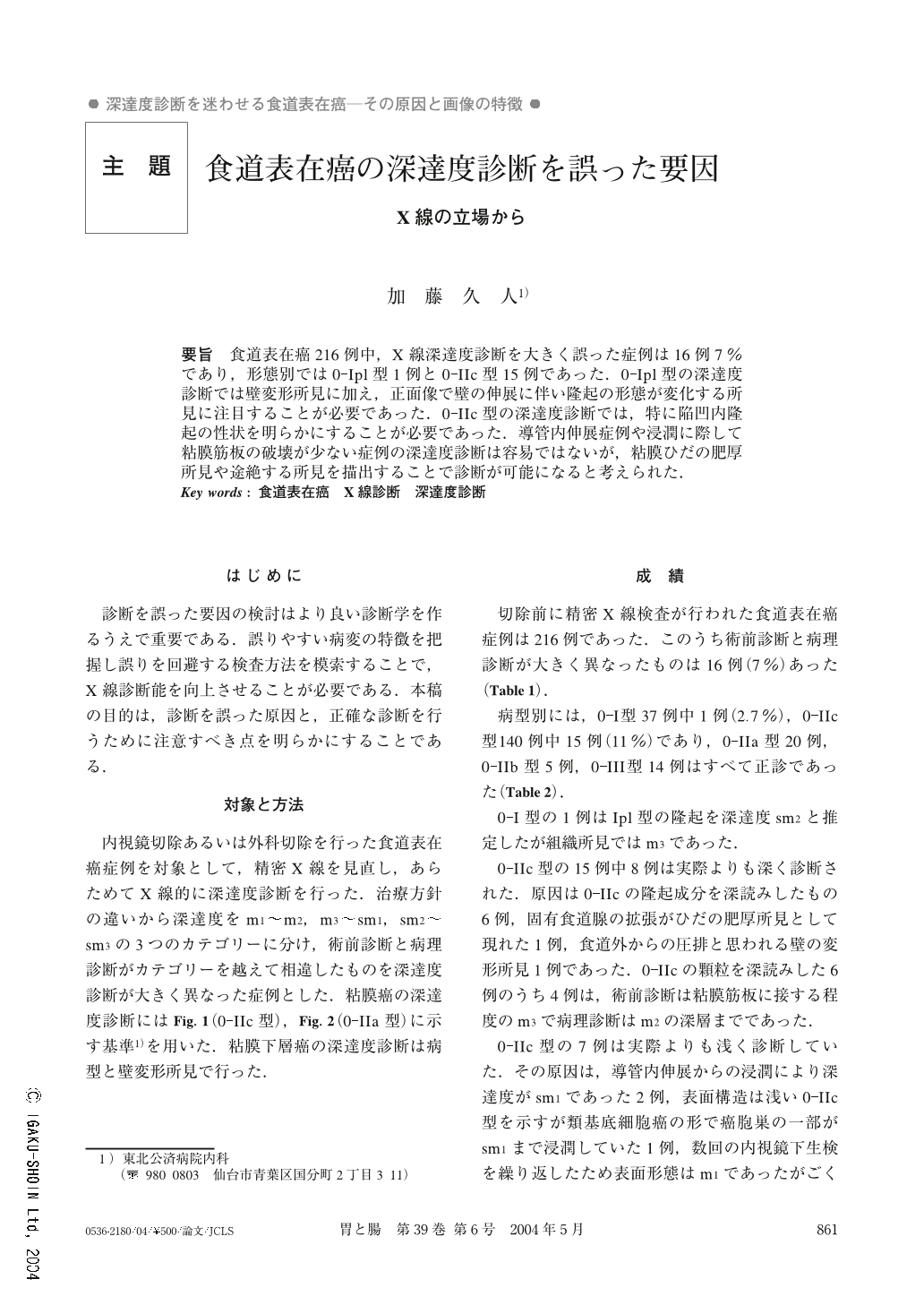
要旨 食道表在癌216例中,X線深達度診断を大きく誤った症例は16例7%であり,形態別では0-Ipl型1例と0-IIc型15例であった.0-Ipl型の深達度診断では壁変形所見に加え,正面像で壁の伸展に伴い隆起の形態が変化する所見に注目することが必要であった.0-IIc型の深達度診断では,特に陥凹内隆起の性状を明らかにすることが必要であった.導管内伸展症例や浸潤に際して粘膜筋板の破壊が少ない症例の深達度診断は容易ではないが,粘膜ひだの肥厚所見や途絶する所見を描出することで診断が可能になると考えられた.
The aim of this study was to clarify causes of errors in radiological estimation of invasion of superficial esophageal cancer and how to avoid them.
Subjects were216cases with superficial esophageal cancer, who underwent radiological examinations before endoscopic or surgical resection. Sixteen cases (7%) with errors of estimation of cancer invasion were noticed. In respect to morphology, they contained one with0-Ipl type and15with0-IIc type. Using radiological diagnosis for0-Ipl type lesions, not only marginal deformities but protrusions stretchable in accord with esophageal distension, are hints leading to a correct diagnosis.
A tip for diagnosis of0-IIc type lesions was to clarify characteristics of granules in the depressed lesions, upward growth or downward growth. It was not easy to estimate cancer invasion from ductal involvement and non-destructive invasion. But it was thought that findings of thickened folds and interrupted folds could be of help for diagnosing of them.
1) Department of Internal Medicine, Tohoku Kousai Hospital, Senndai, Japan
Copyright © 2004, Igaku-Shoin Ltd. All rights reserved.