Japanese
English
- 有料閲覧
- Abstract 文献概要
- 1ページ目 Look Inside
- 参考文献 Reference
- サイト内被引用 Cited by
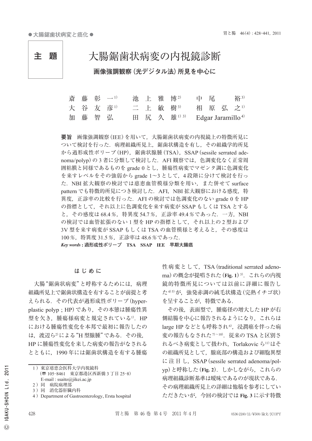
要旨 画像強調観察(IEE)を用いて,大腸鋸歯状病変の内視鏡上の特徴所見について検討を行った.病理組織所見上,鋸歯状構造を有し,その組織学的所見から過形成性ポリープ(HP),鋸歯状腺腫(TSA),SSAP(sessile serrated adenoma/polyp)の3者に分類して検討した.AFI観察では,色調変化なく正常周囲粘膜と同様であるものをgrade 0とし,腫瘍性病変でマゼンタ調に色調変化を来すレベルをその強弱からgrade 1~3として,4段階に分けて検討を行った.NBI拡大観察の検討では慈恵血管模様分類を用い,また併せてsurface patternでも特徴的所見につき検討した.AFI,NBI拡大観察における感度,特異度,正診率の比較を行った.AFIの検討では色調変化のないgrade 0をHPの指標として,それ以上に色調変化を来す病変がSSAPもしくはTSAとすると,その感度は68.4%,特異度54.7%,正診率49.4%であった.一方,NBIの検討では血管拡張のない1型をHPの指標として,それ以上の2型および3V型を来す病変がSSAPもしくはTSAの血管模様と考えると,その感度は100%,特異度31.5%,正診率は48.6%であった.
Seventy-one serrated lesions resected by endoscopic or surgical methods were studied under IEE (image-enhanced endoscopy) including AFI (autofluorescence imaging), NBI (narrow band imaging) and ME (magnifying endoscopy). Serrated lesions were classified into three categories : HP (hyperplastic polyp), SSAP (sessile serrated adenoma/polyp) and TSA (traditional serrated adenoma). Eighteen HPs, 31 SSAPs and 19 TSAs are described. Three serrated lesions with intramucosal cancer (M-Ca) were also studied.
Magenta color changes observed under AFI were graded using a 0-3 scale. Grade 0 with no change while grade 1, grade 2 and grade 3 denoted weak, moderate and strong magenta color changes, respectively.
Capillary-pattern using NBI with ME was classified into four groups (Figure 4). Type 1 with no clear visible capillaries and an unrecognized course ; type 2 with slightly dilated capillaries ; type 3 with markedly dilated capillaries and type 4 with sparse capillaries not following an obvious vascular course. Type 3 pattern was further divided into 2 subtypes : 3V, with a regular course of capillaries observed exclusively in lesions presenting type IV pit pattern along with a villous component and subtype 3I with capillary irregularities such as tortuousness, abrupt caliber change, and heterogeneity in shape.
Additional analysis of the pit pattern under NBI was performed. Three types of pit pattern were observed : Star shaped glands (type II pit), dilated round and/or oval pits (II-D pit) and villous pits (type IV pit). Type II-D pit pattern was observed in 78.9% of SSAPs but only in 35.7% of HPs.
The diagnostic accuracy to differentiate between HP (non-neoplastic lesion) and SSAP or TSA (neoplastic lesion) based on the findings of AFI and NBI was determined prospectively. When grade 0 in AFI observation was selected as indicator of HP, sensitivity and specificity were 68.4% and 54.7% respectively. In contrast, when capillary pattern type 1 in NBI magnifying observation was selected as indicator of HP, sensitivity and specificity were 100% and 31.5% respectively. Accuracy rate as an indicator was not significantly different from AFI diagnosis (49.4%) and NBI diagnosis (48.6%).
Our results suggest that image-enhanced endoscopy might be helpful in the in vivo differentiation of serrated lesions. A new subtype of pit pattern (II-D) is being proposed here. type II-D pit pattern seems to be frequent in SSAPs and its recognition might be of importance to discriminate serrated lesions. Further studies to support our findings are needed.

Copyright © 2011, Igaku-Shoin Ltd. All rights reserved.


