Japanese
English
- 有料閲覧
- Abstract 文献概要
- 1ページ目 Look Inside
- 参考文献 Reference
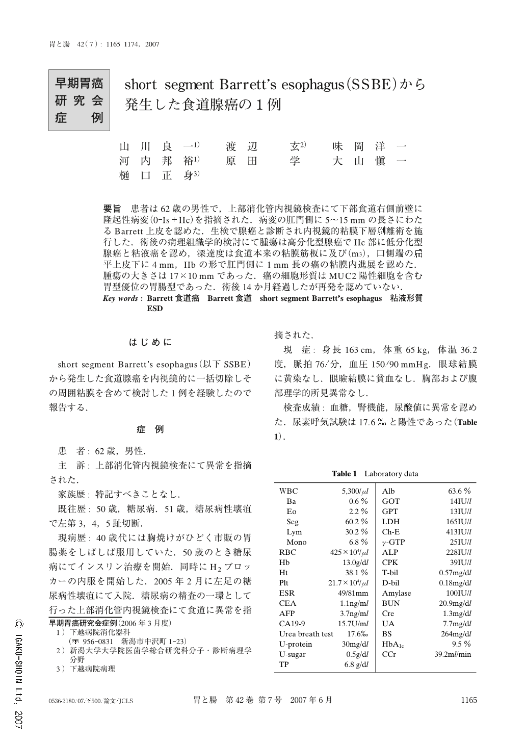
要旨 患者は62歳の男性で,上部消化管内視鏡検査にて下部食道右側前壁に隆起性病変(0-Is+IIc)を指摘された.病変の肛門側に5~15mmの長さにわたるBarrett上皮を認めた.生検で腺癌と診断され内視鏡的粘膜下層剥離術を施行した.術後の病理組織学的検討にて腫瘍は高分化型腺癌でIIc部に低分化型腺癌と粘液癌を認め,深達度は食道本来の粘膜筋板に及び(m3),口側端の扁平上皮下に4mm,IIbの形で肛門側に1mm長の癌の粘膜内進展を認めた.腫瘍の大きさは17×10mmであった.癌の細胞形質はMUC2陽性細胞を含む胃型優位の胃腸型であった.術後14か月経過したが再発を認めていない.
A 62-year-old male underwent upper endoscopic examination. It revealed an elevated lesion (0-Is+IIc) on the right anterior wall of the lower esophagus. Barrett's epithelium 5 to 15mm in length could be seen more on the oral side of the lesion. The biopsy specimens taken from the lesion revealed well-differentiated adenocarcinoma. Endoscopic submucosal dissection (ESD) was performed. Histological study of the resected specimen demonstrated that a well differentiated adenocarcinoma with poorly differentiated and mucinous adenocarcinoma at the IIc area of the tumor, was limited to the esophageal original muscularis mucosae (m3). The cancer extends 4mm in length beneath the non neoplastic squamous epithelium at the oral side of the tumor and 1mm at the anal side in IIb form. The size of the lesion was 17×10mm. Immunostaining revealed that the phenotype of the cancer cells was gastric predominant gastrointestinal type, with MUC 2 positive cells. The patient has survived for 14 months without cancer recurrence after ESD.

Copyright © 2007, Igaku-Shoin Ltd. All rights reserved.


