Japanese
English
- 有料閲覧
PPV購入案内
1,320円 (1,200円+税10%) こちらは、195ページから198ページの文献です。購入には医書.jp本会員登録が必要です。
会員登録完了後に改めて上記「購入サイトへ」を選択してください。
または「購入サイトへ」選択後に「購入ログイン」、「新規会員登録」の順に進んでください。
会員登録について詳しくはをご確認ください。
- Abstract 文献概要
- 1ページ目 Look Inside
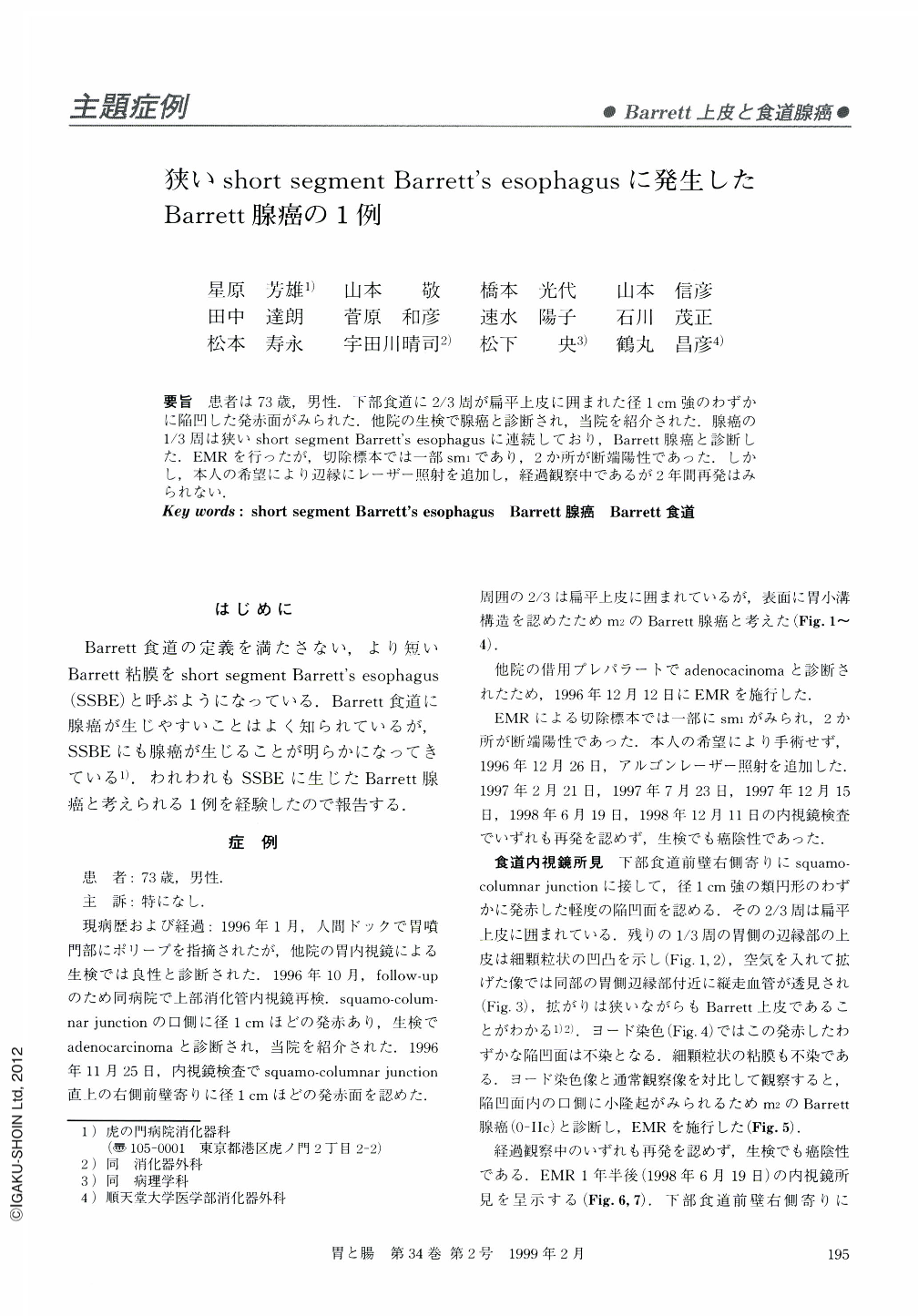
要旨 患者は73歳,男性.下部食道に2/3周が扁平上皮に囲まれた径1cm強のわずかに陥凹した発赤面がみられた.他院の生検で腺癌と診断され,当院を紹介された.腺癌の1/3周は狭いshort segment Barrett's esophagusに連続しており,Barrett腺癌と診断した.EMRを行ったが,切除標本では一部sm1であり,2か所が断端陽性であった.しかし,本人の希望により辺縁にレーザー照射を追加し,経過観察中であるが2年間再発はみられない.
A case of 73-year-old man. A slightly depressed reddish lesion over one centimeter in diameter was detected in the lower esophagus. Two thirds of the lesion was surrounded by squamous mucosa. Biopsy specimens in another hospital revealed adenocarcinoma and the patient was introduced to our hospital. The lesion was diagnosed as Barrett's adenocarcinoma because one third of the lesion extended into the short segment Barrett's esophagus. EMR was performed. The resected specimen revealed that its invasion was partly sm1, and the cut end was positive for carcinoma in two places. Laser irradiation was applied to the surrounding mucosa because the patient rejected an operation. Recurrence hasn't been observed in the two years since the diagnosis.
Copyright © 1999, Igaku-Shoin Ltd. All rights reserved.