雑誌文献を検索します。書籍を検索する際には「書籍検索」を選択してください。

検索

書誌情報 詳細検索 by 医中誌

Japanese

Know-How for Magnifying Observation : Photography, How to Focus, How to Use Different Pigments, Etc.―Mainly the Know-how for Magnifying Endoscopic Observation to Decide Appropriate Therapeutic Strategy Against Neoplastic Colorectal Lesion Hisashi Nakamura 1 , Akihiko Yamamura 2 , Yasuhiro Oono 3 , Yousuke Iriguchi 3 , Jhoji Oda 3 , Masaru Mizutani 3 , Tomoaki Shinohara 3 , Satoshi Takayanagi 3 , Yasuhiro Tomino 3 , Daisuke Kishi 3 , Jun Matsumoto 4 1Department of Gastroenterology, Chofu Surgical Clinic, Chofu, Japan 2Department of Pathology, Tokyo Metropolitan Tama Cancer Detection Center, Fuchu, Japan 3Department of Gastroenterology, Tokyo Metropolitan Tama Cancer Detection Center, Fuchu, Japan 4Department of Surgery, Tokyo Metropolitan Fuchu Hospital, Fuchu, Japan Keyword: 拡大内視鏡 , pit pattern診断 , 色素内視鏡 , 通常内視鏡 , SM深部浸潤癌 pp.645-654
Published Date 2007/4/26
DOI https://doi.org/10.11477/mf.1403101060
  • Abstract
  • Look Inside
  • Reference
  • Cited by

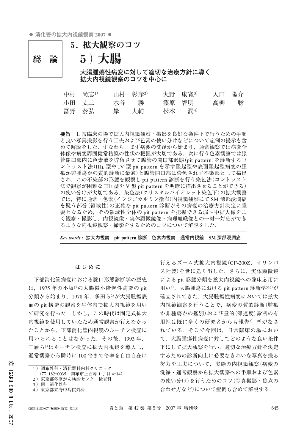
 The procedure for magnifying endoscopic observation and photography under good conditions in daily clinical practice is interpreted through example cases related to ingenuity for good photography and how to use different pigments. Namely, the procedure starts from cleaning the lesion, and in ordinary observation, it is important to grasp the entire picture of the lesion and the status of the peri-lesion normal mucosa. In pigments of observation, it is important to separately use the contrast method in which the gland duct pit pattern is diagnosed by retention of a pigment fluid in the gland duct pit (suitable for qualitative diagnosis of protruding tumors with a pit pattern of Type-IIIL or Type-IV, neoplastic or non-neoplastic) and the staining method in which the gland duct pit is visualized as the non-stained part and the pit pattern is diagnosed by observing the non-stained part (a pit of Type-IIIs or Type V which can hardly be observed by the contrast method can be clearly visualized). In magnifying observation using the staining method (under crystal violet staining), the exact pit pattern diagnosis of the part at which SM deep invasion is suspected (range-type) in conventional colonoscopy or the chromoendoscopy (indigo-carmine spray) is important for deciding the therapeutic strategy against the lesion concerned. There is an explanation of the know-how necessary for the sufficient observation and photography of a low power image enabling a grasp of the pit pattern througout the entire area. In addition there is explanation of the endoscopic observation/photography needed to obtain correspondence between the endoscopic image, stereomicroscopic image and histopathological image.

Copyright © 2007, Igaku-Shoin Ltd. All rights reserved.

基本情報

電子版ISSN 1882-1219 印刷版ISSN 0536-2180 医学書院

関連文献

  • 5)表層拡大型 入口 陽介 , 小田 丈二 , 水谷 勝 , 大野 康寛 , 高柳 聡 , 冨野 泰弘 , 岸 大輔 , 板橋 浩一 , 大村 秀俊 , 篠原 知明 , 藤崎 聡 , 山田 耕三 , 山村 彰彦 , 中村 尚志 , 細井 董三 医学書院 胃と腸
    44巻 4号 (2009年4月)
    pp.553-562
    • 有料閲覧
  • 食道炎のX線診断―逆流性食道炎を中心とする食道・胃接合部における炎症性病変と腫瘍性病変のX線的鑑別診断 小田 丈二 , 入口 陽介 , 水谷 勝 , 高柳 聡 , 冨野 泰弘 , 小山 真一郎 , 岸 大輔 , 藤田 直哉 , 大村 秀俊 , 板橋 浩一 , 中河原 亜希子 , 山里 哲郎 , 山村 彰彦 , 細井 董三 , 秋里 和夫 , 川田 研郎 , 西蔭 徹郎 , 河野 辰幸 , 今村 和弘 , 小林 里絵 , 松本 潤 医学書院 胃と腸
    46巻 8号 (2011年7月)
    pp.1178-1193
    • 有料閲覧
  • linitis plastica型胃癌の自然史 入口 陽介 , 細井 董三 , 小田 丈二 , 水谷 勝 , 大野 康寛 , 篠原 知明 , 高柳 聡 , 冨野 泰弘 , 岸 大輔 , 大村 秀俊 , 霧生 信昭 , 細井 亜希子 , 藤崎 聡 , 板橋 浩一 , 高倉 裕一 , 大浦 通久 , 山田 耕三 , 中村 尚志 , 中橋 栄太 , 山村 彰彦 医学書院 胃と腸
    43巻 5号 (2008年4月)
    pp.751-763
    • 有料閲覧
  • 肛門管尖圭コンジローマの1例 大野 康寛 , 入口 陽介 , 山村 彰彦 , 小田 丈二 , 水谷 勝 , 岸 大輔 , 大浦 通久 , 冨野 泰弘 , 高柳 聡 , 藤崎 聡 , 篠原 知明 , 山田 耕三 , 池松 弘朗 , 中村 尚志 , 細井 董三 医学書院 胃と腸
    45巻 10号 (2010年9月)
    pp.1699-1706
    • 有料閲覧
  • 内視鏡経過例における早期胃癌発育の遡及的検討―粘液形質からみて 入口 陽介 , 小田 丈二 , 水谷 勝 , 大野 康寛 , 高柳 聡 , 篠原 知明 , 富野 泰弘 , 岸 大輔 , 藤崎 聡 , 大村 秀俊 , 板橋 浩一 , 山田 耕三 , 中村 尚志 , 山村 彰彦 , 細井 董三 医学書院 胃と腸
    43巻 12号 (2008年11月)
    pp.1752-1763
    • 有料閲覧
  • 食道胃接合部腺癌のX線診断―早期癌形態を呈した病変の臨床病理学的特徴から 小田 丈二 , 入口 陽介 , 水谷 勝 , 大野 康寛 , 高柳 聡 , 冨野 泰弘 , 藤崎 聡 , 篠原 知明 , 岸 大輔 , 大村 秀俊 , 板橋 浩一 , 細井 亜希子 , 山田 耕三 , 細井 董三 , 山村 彰彦 医学書院 胃と腸
    44巻 7号 (2009年6月)
    pp.1128-1143
    • 有料閲覧
  • 微小胃癌に対するX線診断の現状と課題 入口 陽介 , 小田 丈二 , 水谷 勝 , 高柳 聡 , 冨野 泰弘 , 小山 真一郎 , 山里 哲郎 , 岸 大輔 , 大村 秀俊 , 板橋 浩一 , 中河原 亜希子 , 藤田 直哉 , 今村 和広 , 高西 喜重郎 , 松本 潤 , 山村 彰彦 , 細井 董三 医学書院 胃と腸
    48巻 6号 (2013年5月)
    pp.822-834
    • 有料閲覧
  • 過形成,鋸歯状腺腫から発育進展した大腸低分化腺癌の1例 冨野 泰弘 , 入口 陽介 , 小田 丈二 , 水谷 勝 , 高柳 聡 , 岸 大輔 , 小山 真一郎 , 板橋 浩一 , 藤田 直哉 , 細井 亜希子 , 大村 秀俊 , 北條 裕美子 , 山田 耕三 , 今村 和広 , 足立 健介 , 松本 潤 , 山村 彰彦 , 細井 董三 医学書院 胃と腸
    45巻 11号 (2010年10月)
    pp.1808-1814
    • 有料閲覧
  • 多発胃癌の診断におけるX線造影検査の役割 入口 陽介 , 小田 丈二 , 水谷 勝 , 高柳 聡 , 冨野 泰弘 , 小山 真一郎 , 岸 大輔 , 板橋 浩一 , 大村 秀俊 , 藤田 直哉 , 細井 亜希子 , 北條 裕美子 , 山田 耕三 , 今村 和広 , 松本 潤 , 山村 彰彦 , 細井 董三 医学書院 胃と腸
    46巻 1号 (2011年1月)
    pp.31-45
    • 有料閲覧
  • 低異型度分化型胃癌のX線像の検討 入口 陽介 , 小田 丈二 , 水谷 勝 , 高柳 聡 , 冨野 泰弘 , 小山 真一郎 , 岸 大輔 , 大村 秀俊 , 板橋 浩一 , 藤田 直哉 , 北條 裕美子 , 細井 亜希子 , 山田 耕三 , 今村 和弘 , 松本 潤 , 山村 彰彦 , 細井 董三 医学書院 胃と腸
    45巻 7号 (2010年6月)
    pp.1097-1113
    • 有料閲覧
もっと見る

文献を共有