Japanese
English
- 有料閲覧
- Abstract 文献概要
- 1ページ目 Look Inside
- 参考文献 Reference
- サイト内被引用 Cited by
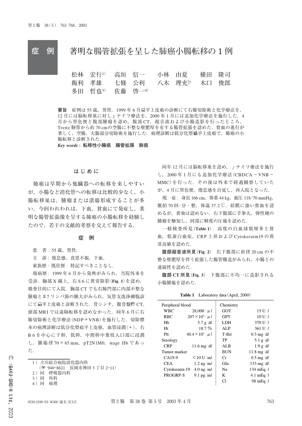
要旨 症例は55歳,男性.1999年6月扁平上皮癌の診断にて右肺切除術と化学療法を,12月には脳転移巣に対しγナイフ療法を,2000年1月には追加化学療法を施行した.4月から黒色便と腹部腫瘤を認め,腹部CT,超音波および小腸造影を行ったところ,Treitz靭帯から約70cmの空腸に不整な壁肥厚を有する腸管拡張を認めた.貧血の進行が著しく,空腸,大腸部分切除術を施行した.病理診断は低分化型扁平上皮癌で,肺癌の小腸転移と診断された.
In the literature survey, most of the mestastases of lung carcinomas macroscopically from tumor and/or ulcer. The size is under 10 cm in the small intestine, and they manifest with perforation, ileus, invagination or bloody discharge. We reported a 55-year-old man having poorly-differentiated pulmonary squamous cell carcinoma which resulted in jejunal metastasis and formed an obvious luminal dilatation, sized lung 20 cm. The patient underwent right pneumonectomy and the clinical stage of the lung cancer was IIb (pT2N1M0). Six months after, γ-knife therapy and systemic chemotherapy (CBDCA+VNB+MMC) was carried out for brain metastasis. Ten months after the lung operation. The patient was admitted with severe anemia and bloody bowel discharge. We examined him using US, CT and contrast medium of the small intestine, and the tumor was shown to have formed obvious luminal dilatation with an irregularly thickened wall, 70 cm anal from the Treitz ligament. We performed partial jejunectomy and transverse colonectomy, but the patient died of carcinomatosa peritonitis one month after the operation. In the resected material, the tumor was found to be mainly located in the subserosa with many venous permeations. We considered the tumor to have progressed mainly in the subserosa, so that the metastasis formed a large dilatation and manifested itself after it reached a size as large as 20 cm. We recommended careful follow-up of pulmonary carcinoma patients using fecal occult blood tests and image analysis after pneumonectomy.

Copyright © 2003, Igaku-Shoin Ltd. All rights reserved.


