Japanese
English
- 有料閲覧
- Abstract 文献概要
- 1ページ目 Look Inside
- 参考文献 Reference
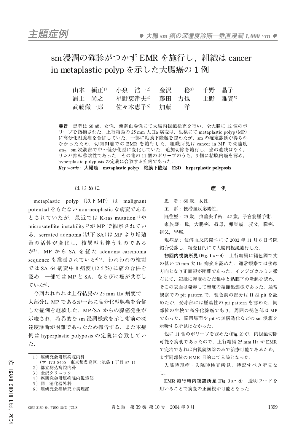
要旨 患者は60歳,女性.便潜血陽性にて大腸内視鏡検査を行い,全大腸に12個のポリープを指摘された.上行結腸の25mm大IIa病変は,生検にてmetaplastic polyp(MP)に高分化型腺癌を合併していた.一部に粘膜下隆起を認めたが,smの確定診断が得られなかったため,切開剥離でのEMRを施行した.組織所見はcancer in MPで深達度sm2,sm浸潤部で中~低分化型に変化していた.追加切除を施行し,癌の遺残はなく,リンパ節転移陰性であった.その他の11個のポリープのうち,3個に粘膜内癌を認め,hyperplastic polyposisの定義に合致する症例であった.
A 60-year-old woman underwent colonoscopic examination for faecal occult blood. There were 12 polyps in her large intestine.
A flat lesion 25 mmin diameter in the ascending colon was composed of two parts. Biopsy specimens demonstrate that the very flat part of the lesion is metaplastic polyp, and the protruded part of that is well-differentiated adenocarcinama.
We performed endoscopical submucosal dissection in order to resect the lesion en bloc.
The pathological diagnosis of the lesion was carcinoma in a metaplastic polyp. The carcinoma had invaded the submucosal layer to the depth of 1,700μm. It was re-evaluated to moderate-to-poorly differentiated adenocarcinoma at the invading front. Ileocecal resection was perfomed and there was no residual carcinoma or lymphnode metastasis. In addition, 3 of 11 polyps endoscopically resected contained intramucosal carcinoma. This patient can be categorized as a case of hyperplastic polyposis.
1) Department of Internal Medicine, Cancer Institute Hospital, Tokyo
2) Department of Internal Medicine, Tokyo Metropolitan Komagome Hospital, Tokyo

Copyright © 2004, Igaku-Shoin Ltd. All rights reserved.


