Japanese
English
- 有料閲覧
- Abstract 文献概要
- 1ページ目 Look Inside
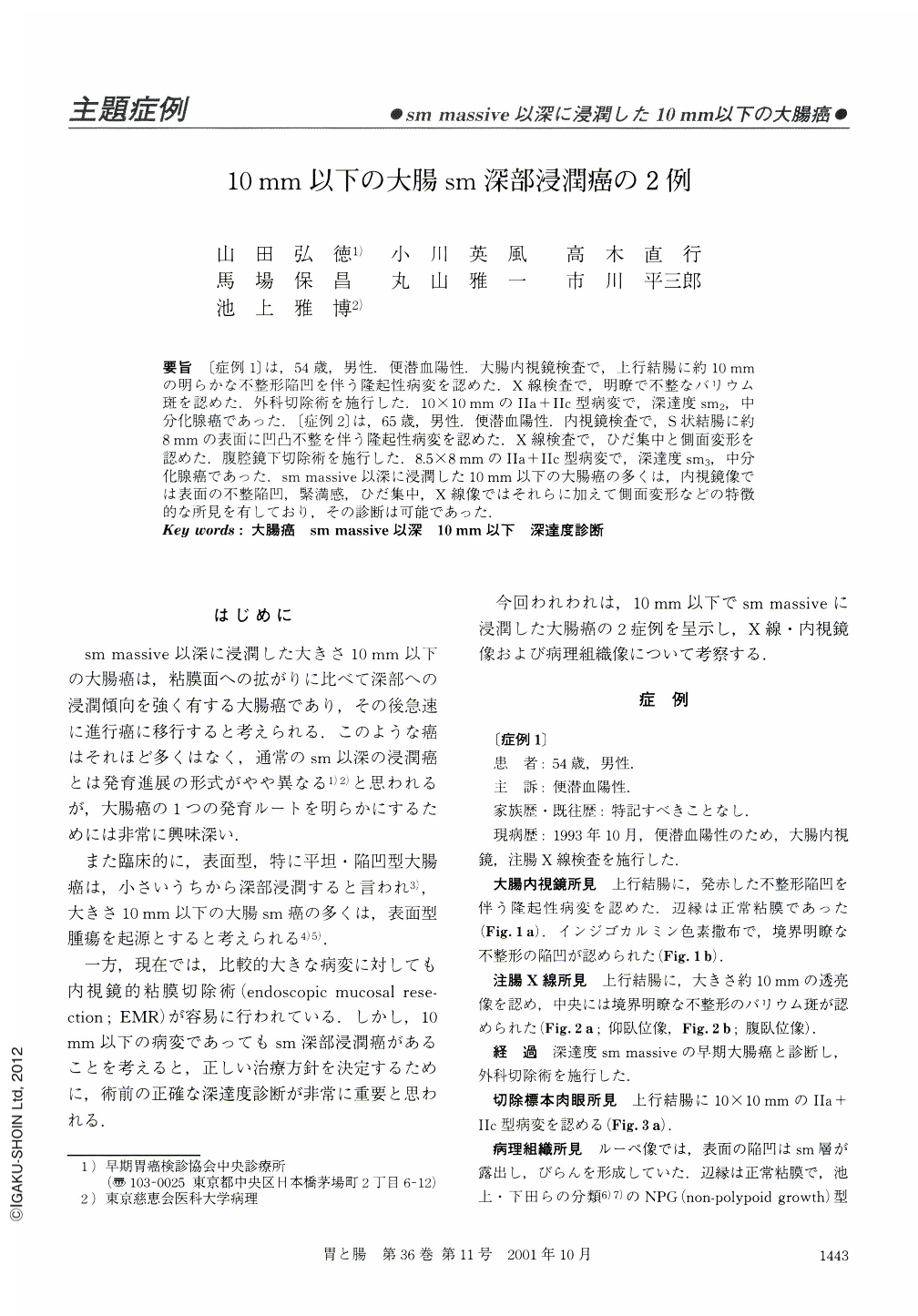
要旨 〔症例1〕は,54歳,男性.便潜血陽性.大腸内視鏡検査で,上行結腸に約10mmの明らかな不整形陥凹を伴う隆起性病変を認めた.X線検査で,明瞭で不整なバリウム斑を認めた.外科切除術を施行した.10×10mmのⅡa+Ⅱc型病変で,深達度sm2,中分化腺癌であった.〔症例2〕は,65歳,男性.便潜血陽性.内視鏡検査で,S状結腸に約8mmの表面に凹凸不整を伴う隆起性病変を認めた.X線検査で,ひだ集中と側面変形を認めた.腹腔鏡下切除術を施行した.8.5×8mmのⅡa+Ⅱc型病変で,深達度sm3,中分化腺癌であった.sm massive以深に浸潤した10mm以下の大腸癌の多くは,内視鏡像では表面の不整陥凹,緊満感,ひだ集中,X線像ではそれらに加えて側面変形などの特徴的な所見を有しており,その診断は可能であった.
〔Case 1〕: A 54-year-old, male; Colonoscopy revealed a flat-elevated lesion of the ascending colon with central depression. Double contrast barium enema revealed the central depression distinctly. Surgical resection of the colon was performed. Macroscopic findings showed a IIa+IIc-type lesion, measuring 10×10 mm in the ascending colon. Histologically, it was shown that moderately differentiated adenocarcinoma had invaded the submucosal layer with lymphatic permeation (sm2, ly1, v0, n0).
〔Case 2〕: A 65-year-old, male; Colonoscopy revealed a flat-elevated lesion with irregular surface in the sigmoid colon. Double contrast barium enema revealed a depression and the basal indentation of the lesion. Laparoscopic sigmoidectomy was performed. Macroscopic findings showed a IIa+IIc-type lesion, measuring 8.5×8 mm in the sigmoid colon. Histologically, it was shown that moderately differentiated adenocarcinoma had deeply invaded the submucosal layer with vessel invasion (sm3, ly0, v1, n0). Colorectal carcinomas, less than 10 mm in diameter, with massive submucosal invasion, could thus be diagnosed without difficulty based on meticulous analysis of the colonoscopic and radiographic images.

Copyright © 2001, Igaku-Shoin Ltd. All rights reserved.


