Japanese
English
- 有料閲覧
PPV購入案内
1,320円 (1,200円+税10%) こちらは、1455ページから1460ページの文献です。購入には医書.jp本会員登録が必要です。
会員登録完了後に改めて上記「購入サイトへ」を選択してください。
または「購入サイトへ」選択後に「購入ログイン」、「新規会員登録」の順に進んでください。
会員登録について詳しくはをご確認ください。
- Abstract 文献概要
- 1ページ目 Look Inside
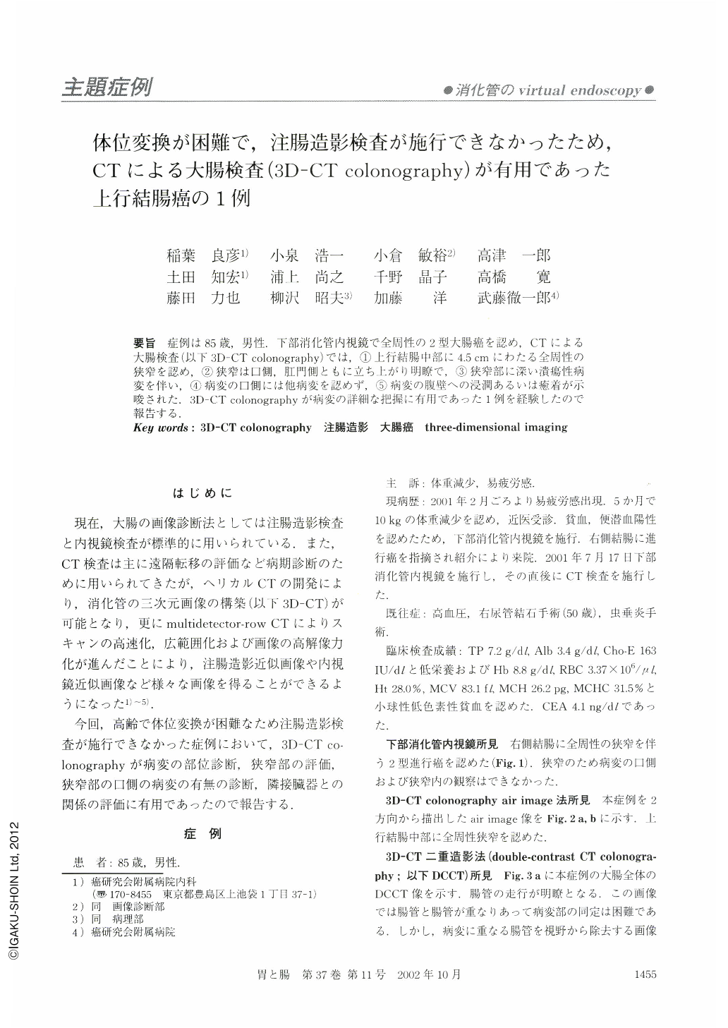
要旨 症例は85歳,男性.下部消化管内視鏡で全周性の2型大腸癌を認め,CTによる大腸検査(以下3D-CT colonography)では,①上行結腸中部に4.5cmにわたる全周性の狭窄を認め,②狭窄は口側,肛門側ともに立ち上がり明瞭で,③狭窄部に深い潰瘍性病変を伴い,④病変の口側には他病変を認めず,⑤病変の腹壁への浸潤あるいは癒着が示唆された.3D-CT colonographyが病変の詳細な把握に有用であった1例を経験したので報告する.
An 85-year-old man suffering from colonic cancer, visited the Cancer Institute Hospital, because of anemia. Endoscopic examination disclosed annular advanced colonic cancer with stenosis in the right side colon. 3D-CT colonography showed stenosis with a deep ulceration, 4.5 cm in length, in the ascending colon. There was no obvious lesion at the oral side of the cancer. In addition, minimal invasion or adhesion to the abdominal wall was suspected. Surgical specimen showed annular carcinoma adhering to the abdominal wall.
3D-CT colonography is a good means for assessing what stage a colonic cancer is in, before undertaking a surgical operation.
Copyright © 2002, Igaku-Shoin Ltd. All rights reserved.