Japanese
English
- 有料閲覧
- Abstract 文献概要
- 1ページ目 Look Inside
- 参考文献 Reference
- サイト内被引用 Cited by
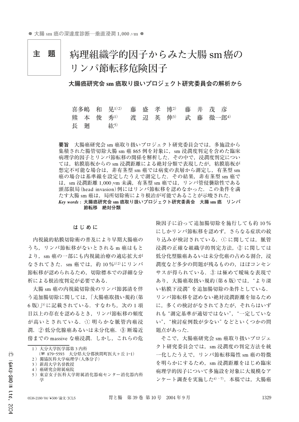
要旨 大腸癌研究会sm癌取り扱いプロジェクト研究委員会では,多施設から集積された腸管切除大腸sm癌865例を対象に,sm浸潤度判定を含めた臨床病理学的因子とリンパ節転移の関係を解析した.その中で,浸潤度判定については,粘膜筋板からのsm浸潤距離による絶対分類で表現したが,粘膜筋板が想定不可能な場合は,非有茎型sm癌では病変の表層から測定し,有茎型sm癌の場合は基準線を設定したうえで測定した.その結果,非有茎型sm癌では,sm浸潤距離1,000μm未満,有茎型sm癌では,リンパ管侵襲陰性である頭部限局(head invasion)例にはリンパ節転移を認めなかった.この条件を満たす大腸sm癌は,局所切除術により根治が可能であることが示唆された.
The Japanese Society for Cancer of the Colon and Rectum analyzed Clinicopathological factors of865submucosal invasive colorectal carcinomas, from865patients who had undergone surgical resection at six institutions.
In measuring the depth of submucosal invasion, submucosal invasive colorectal carcinomas were classified into two types, pedenculated and non-superficial types.
When the muscularis mucosae could be identified in HE or Desmin stained specimens, the muscularis mucosae was used as baseline and the vertical distance from this line to the deepest portion of invasion represented the depth of submucosal invasion.
When the muscularis mucosae could not be identified due to carcinomatous invasion, we determine the baseline according to the macroscopic types.
For pedunculated SICC, a border between normal epithelium and neoplastic epithelium was used as baseline and the depth of submucosal invasion was measured as the vertical distance from this line to the deepest portion of invasion. When the deepest portion of invasion was limited to above the baseline, the case was defined as a head invasion and SM depth was regarded as0μm. When the deepest portion of invasion was located below the baseline, the case was defined as a stalk invasion and the vertical distance from this line to the deepest portion of invasion was the depth of submucosal invasion.
For non-pedunculated SICC, the tumor surface was used as baseline, and the vertical distance from this line to the deepest portion of invasion represented the depth of submucosal invasion.
For pedunculated SICC, the rate of lymph-node metastasis was0% in head invasion cases and stalk invasion cases with a depth of submucosal invasion<3,000μmif lymphatic invasion was negative. For non-pedunculated SICC, the rate of lymph-node metastasis was also0% if the depth of submucosal invasion was <1,000μm.
1) The Third Department of Internal Medicine, Oita University Faculty of Medicine, Oita, Japan
2) Department of Surgical and Molecular Pathology, Dokkyo University School of Medicine, Tochigi, Japan
3) Division of Molecular and Functional Pathology, Department of Cellular Function, Graduate School of Medical and Dental Sciences, Niigata University, Niigata, Japan

Copyright © 2004, Igaku-Shoin Ltd. All rights reserved.


