Japanese
English
- 有料閲覧
- Abstract 文献概要
- 1ページ目 Look Inside
- 参考文献 Reference
- サイト内被引用 Cited by
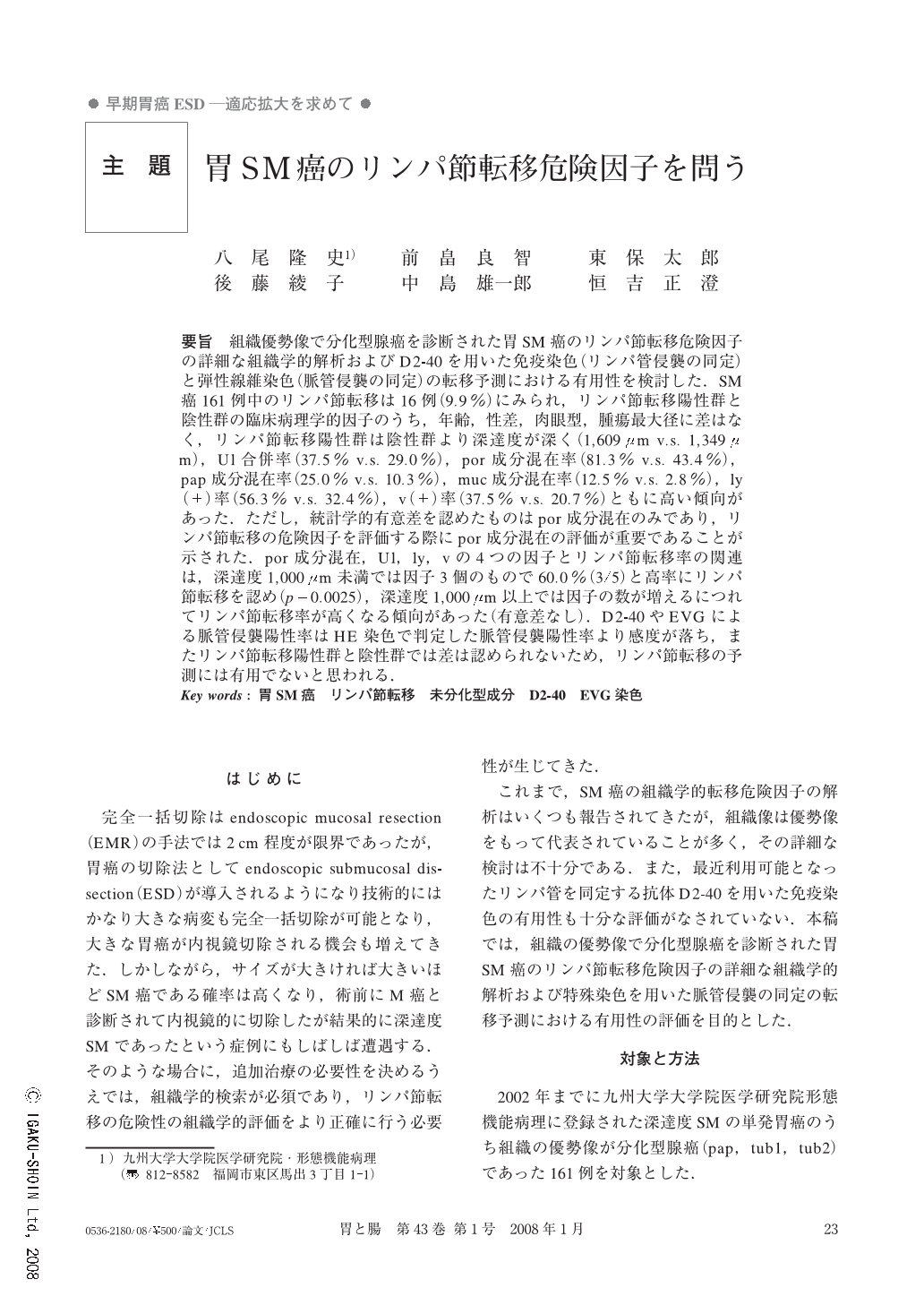
要旨 組織優勢像で分化型腺癌を診断された胃SM癌のリンパ節転移危険因子の詳細な組織学的解析およびD2-40を用いた免疫染色(リンパ管侵襲の同定)と弾性線維染色(脈管侵襲の同定)の転移予測における有用性を検討した.SM癌161例中のリンパ節転移は16例(9.9%)にみられ,リンパ節転移陽性群と陰性群の臨床病理学的因子のうち,年齢,性差,肉眼型,腫瘍最大径に差はなく,リンパ節転移陽性群は陰性群より深達度が深く(1,609μm v.s. 1,349μm),Ul合併率(37.5% v.s. 29.0%),por成分混在率(81.3% v.s. 43.4%),pap成分混在率(25.0% v.s. 10.3%),muc成分混在率(12.5% v.s. 2.8%),ly(+)率(56.3% v.s. 32.4%),v(+)率(37.5% v.s. 20.7%)ともに高い傾向があった.ただし,統計学的有意差を認めたものはpor成分混在のみであり,リンパ節転移の危険因子を評価する際にpor成分混在の評価が重要であることが示された.por成分混在,Ul,ly,vの4つの因子とリンパ節転移率の関連は,深達度1,000μm未満では因子3個のもので60.0%(3/5)と高率にリンパ節転移を認め(p=0.0025),深達度1,000μm以上では因子の数が増えるにつれてリンパ節転移率が高くなる傾向があった(有意差なし).D2-40やEVGによる脈管侵襲陽性率はHE染色で判定した脈管侵襲陽性率より感度が落ち,またリンパ節転移陽性群と陰性群では差は認められないため,リンパ節転移の予測には有用でないと思われる.
We evaluated risk factors of lymph node metastasis from gastric adenocarcinomas of predominantly differentiated type, including lymphovascular invasion detected by immunohistochemical stain for D2-40 and special stain for elastic fibers.
Among 161 cases of gastric adenocarcinomas with submucosal invasion, 16 cases (9.9%) had lymph node metastasis. There was no difference due to patient's age, sex ratio, growth type and tumor size between cases with lymph node metastasis (+) and (-). Cases with lymph node metastasis tended to experience deeper invasion (1,609μm v.s. 1,349μm) and a higher incidence of co-existence of ulcers (37.5% v.s. 29.0%), co-existence of poorly differentiated components (81.3% v.s. 43.4%), papillary components (25.0% v.s. 10.3%), mucinous components (12.5% v.s. 2.8%), lymphatic permeation (56.3% v.s. 32.4%) and venous invasion (37.5% v.s. 20.7%). However, only incidence of co-existence of a poorly differentiated component is statistically significant. This result indicates the importance of evaluating the co-existence of a poorly differentiated component, when the risk of lymph node metastasis is evaluated.
Risk of lymph node metastasis was also evaluated with reference to its combination with 4 factors (co-existence of poorly differentiated component, co-existence of ulcer, lymphatic permeation and venous invasion). In the group with depth of invasion less than 1,000μm, the group with three of those factors experienced a higher incidence of lymph node metastasis (60.0%:3/5) than other groups (p=0.0025). In the group with depth of invasion more than 1,000μm, the group with the more factors tended to experience a higher incidence of lymph node metastasis, but this was not statistically significant. Evaluation by special stains (D2-40 and EVG) reduced the sensitivity compared with that by HE stain and was not considered to be useful for improving the prediction of lymph node metastasis.

Copyright © 2008, Igaku-Shoin Ltd. All rights reserved.


