Japanese
English
- 有料閲覧
- Abstract 文献概要
- 1ページ目 Look Inside
- 参考文献 Reference
はじめに
側頭骨内髄膜脳瘤は頭蓋底骨欠損部から頭蓋内容物が頭蓋外へ脱出する病態であり,大半は後頭部に発生し側頭骨内に発生するのは稀である1)。しかし,その症例報告は本邦でも散見され,日常の耳鼻咽喉科診療で遭遇する可能性は十分にある。先天性のもののほか,耳科手術,外傷,中耳感染が成因となりうるが特発性の発生も報告されている2)。耳鼻咽喉科には耳漏を主訴に受診することが多いが,滲出性中耳炎と診断され発見が遅れる場合も多い。
今回,対側の真珠腫性中耳炎の既往や鼓膜,画像所見から天蓋の菲薄化した真珠腫性中耳炎を疑い経乳突洞的に手術を行ったところ,術中の所見および術後病理診断から側頭骨内髄膜脳瘤の診断に至った1例を経験した。過去の文献的考察を踏まえて考察する。
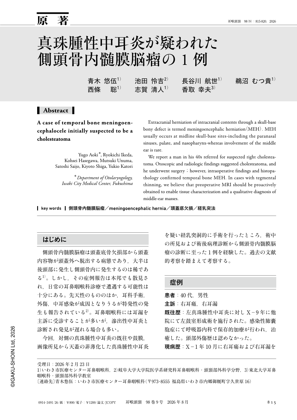
Extracranial herniation of intracranial contents through a skull-base bony defect is termed meningoencephalic herniation(MEH). MEH usually occurs at midline skull-base sites-including the paranasal sinuses, palate, and nasopharynx-whereas involvement of the middle ear is rare.
We report a man in his 40s referred for suspected right cholesteatoma. Otoscopic and radiologic findings suggested cholesteatoma, and he underwent surgery;however, intraoperative findings and histopathology confirmed temporal bone MEH. In cases with tegmental thinning, we believe that preoperative MRI should be proactively obtained to enable tissue characterization and a qualitative diagnosis of middle-ear masses.
Copyright © 2026, Igaku-Shoin Ltd. All rights reserved.