Japanese
English
- 有料閲覧
PPV購入案内
1,650円 (1,500円+税10%) こちらは、407ページから412ページの文献です。購入には医書.jp本会員登録が必要です。
会員登録完了後に改めて上記「購入サイトへ」を選択してください。
または「購入サイトへ」選択後に「購入ログイン」、「新規会員登録」の順に進んでください。
会員登録について詳しくはをご確認ください。
- Abstract 文献概要
- 1ページ目 Look Inside
- 参考文献 Reference
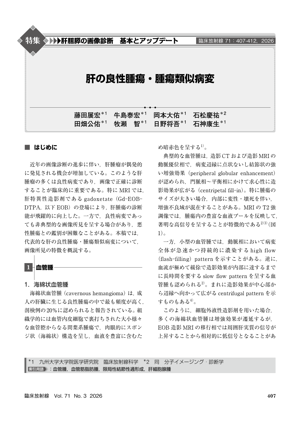
近年の画像診断の進歩に伴い,肝腫瘤が偶発的に発見される機会が増加している。このような肝腫瘤の多くは良性病変であり,画像で正確に診断することが臨床的に重要である。特にMRIでは,肝特異性造影剤であるgadoxetate(Gd-EOB-DTPA,以下EOB)の登場により,肝腫瘍の診断能が飛躍的に向上した。一方で,良性病変であっても非典型的な画像所見を呈する場合があり,悪性腫瘍との鑑別が困難なことがある。本稿では,代表的な肝の良性腫瘍・腫瘍類似病変について,画像所見の特徴を概説する。
With the advancement of imaging modalities, the incidental detection of hepatic lesions has increased. Accurate diagnosis is clinically crucial to differentiate benign entities from malignancies. This article provides a comprehensive overview of the imaging characteristics of benign hepatic tumors and tumor-like lesions, including cavernous hemangioma, infantile hemangioma, angiomyolipoma, focal nodular hyperplasia(FNH), and hepatocellular adenoma(HCA). Understanding the specific imaging patterns and potential diagnostic pitfalls of these lesions is essential for radiologists to provide accurate diagnoses and guide appropriate clinical management.
Copyright © 2026, KANEHARA SHUPPAN Co.LTD. All rights reserved.