Japanese
English
- 有料閲覧
PPV購入案内
1,650円 (1,500円+税10%) こちらは、415ページから418ページの文献です。購入には医書.jp本会員登録が必要です。
会員登録完了後に改めて上記「購入サイトへ」を選択してください。
または「購入サイトへ」選択後に「購入ログイン」、「新規会員登録」の順に進んでください。
会員登録について詳しくはをご確認ください。
- Abstract 文献概要
- 1ページ目 Look Inside
- 参考文献 Reference
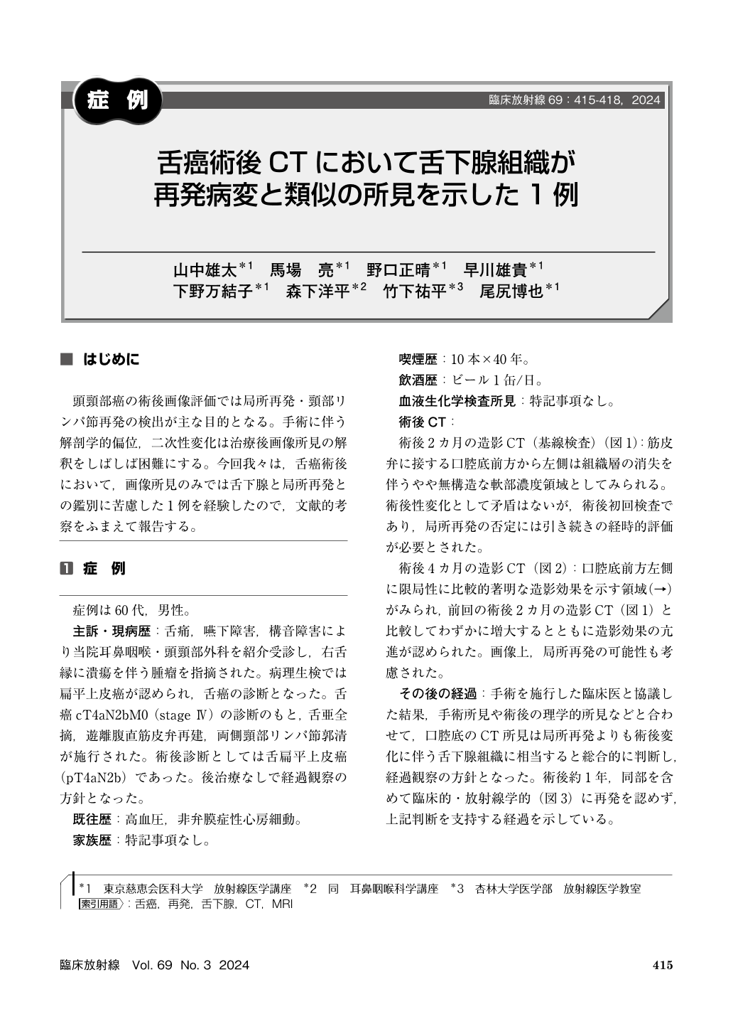
頭頸部癌の術後画像評価では局所再発・頸部リンパ節再発の検出が主な目的となる。手術に伴う解剖学的偏位,二次性変化は治療後画像所見の解釈をしばしば困難にする。今回我々は,舌癌術後において,画像所見のみでは舌下腺と局所再発との鑑別に苦慮した1例を経験したので,文献的考察をふまえて報告する。
A male in his 60s with postoperative status of tongue cancer. On follow–up postoperative serial CTs, a localized, some marked contrast enhancement was noted on the anterior and left side of the oral floor adjacent to the cutaneous flap, which slightly increased in size and showed increased contrast enhancement. Local recurrence could not be ruled out based on the imaging findings alone, and after discussion with the surgeon, it was determined that the anatomic and inflammatory changes in the sublingual gland caused by the surgical procedure. No recurrence was clinically and radiologically detected 1 year later. When it is difficult to distinguish between local recurrence after head and neck cancer surgery and normal tissue following surgery, it is important to discuss the case with the clinician.
Copyright © 2024, KANEHARA SHUPPAN Co.LTD. All rights reserved.