Japanese
English
症例
子宮発生腺肉腫の1例—術前PET/MRI画像と病理標本との対比
A case of uterine adenosarcoma:comparison between PET/MRI and pathological findings
山本 雄也
1,2
,
上野 嘉子
1
,
松田 耕平
3
,
野上 宗伸
1
,
出口 雅士
4
,
上原 慶一郎
5
,
三木 智子
1
,
村上 卓道
1
Katsuya Yamamoto
1,2
1神戸大学大学院医学研究科 内科系講座放射線診断学分野
2兵庫県立丹波医療センター 放射線科
3高槻病院 放射線診断科
4神戸大学大学院医学研究科 外科系講座産科婦人科学分野
5同 病理部学講座病理診断学分野
1Department of Radiology Tamba Medical Center/Kobe University
キーワード:
adenosarcoma
,
uterine
,
PET/MRI
Keyword:
adenosarcoma
,
uterine
,
PET/MRI
pp.381-385
発行日 2021年4月10日
Published Date 2021/4/10
DOI https://doi.org/10.18888/rp.0000001573
- 有料閲覧
PPV購入案内
1,650円 (1,500円+税10%) こちらは、381ページから385ページの文献です。
PPV(ペイ・パー・ビュー)とは、論文(記事)単位で購入・閲覧ができるサービスです。
購入には医書.jp本会員登録が必要です。
会員登録完了後に改めて上記「購入サイトへ」を選択してください。
または「購入サイトへ」選択後に「購入ログイン」、「新規会員登録」の順に進んでください。
会員登録について詳しくはをご確認ください。
購入には医書.jp本会員登録が必要です。
会員登録完了後に改めて上記「購入サイトへ」を選択してください。
または「購入サイトへ」選択後に「購入ログイン」、「新規会員登録」の順に進んでください。
会員登録について詳しくはをご確認ください。
- Abstract 文献概要
- 1ページ目 Look Inside
- 参考文献 Reference
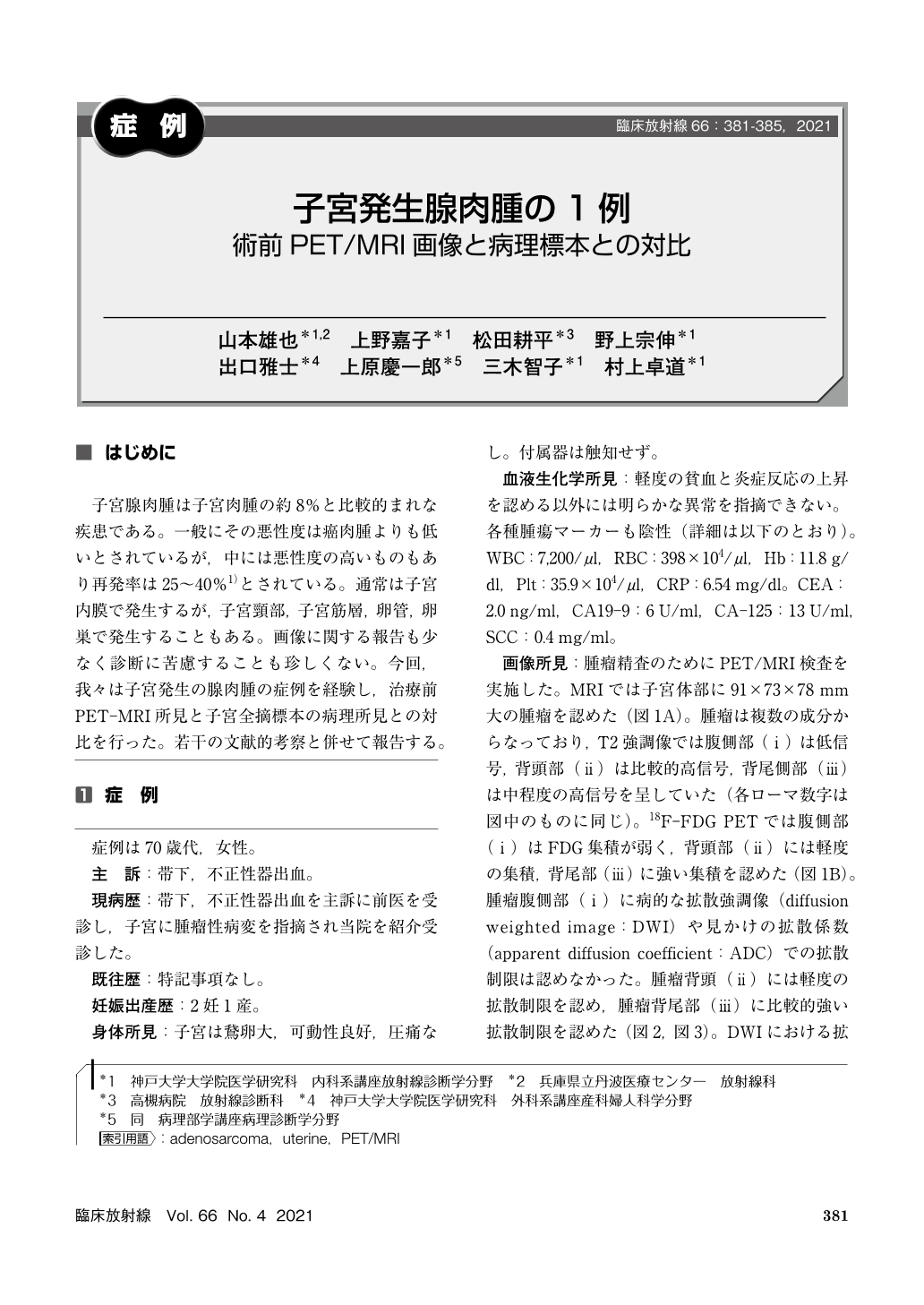
子宮腺肉腫は子宮肉腫の約8%と比較的まれな疾患である。一般にその悪性度は癌肉腫よりも低いとされているが,中には悪性度の高いものもあり再発率は25〜40%1)とされている。通常は子宮内膜で発生するが,子宮頸部,子宮筋層,卵管,卵巣で発生することもある。画像に関する報告も少なく診断に苦慮することも珍しくない。今回,我々は子宮発生の腺肉腫の症例を経験し,治療前PET-MRI所見と子宮全摘標本の病理所見との対比を行った。若干の文献的考察と併せて報告する。
Uterine adenosarcoma is a mixed tumor with a mixture of benign gland epithelium and sarcoma components. We report a case that occurred in a patient in her 70 s. In this case, the invasion into the muscular layer was evident and preoperative diagnosis was difficult. In addition, this disease has benign glandular epithelium, and it sometimes is difficult to diagnose with biopsy specimens. FDG accumulation and diffusion restriction demonstrated by PET/MRI were useful for diagnosis.
Copyright © 2021, KANEHARA SHUPPAN Co.LTD. All rights reserved.