Japanese
English
- 有料閲覧
PPV購入案内
1,650円 (1,500円+税10%) こちらは、1407ページから1410ページの文献です。購入には医書.jp本会員登録が必要です。
会員登録完了後に改めて上記「購入サイトへ」を選択してください。
または「購入サイトへ」選択後に「購入ログイン」、「新規会員登録」の順に進んでください。
会員登録について詳しくはをご確認ください。
- Abstract 文献概要
- 1ページ目 Look Inside
- 参考文献 Reference
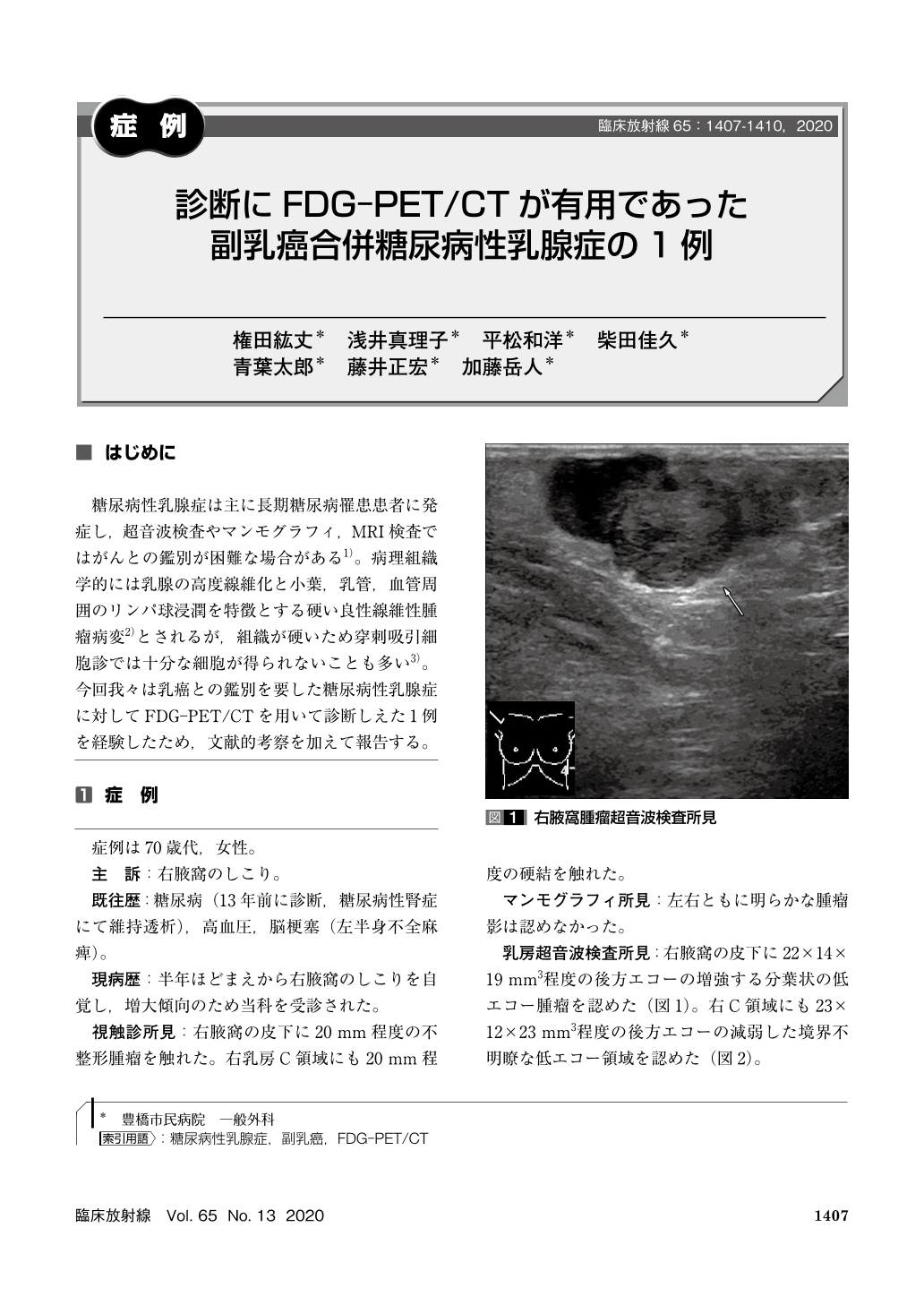
糖尿病性乳腺症は主に長期糖尿病罹患患者に発症し,超音波検査やマンモグラフィ,MRI検査ではがんとの鑑別が困難な場合がある1)。病理組織学的には乳腺の高度線維化と小葉,乳管,血管周囲のリンパ球浸潤を特徴とする硬い良性線維性腫瘤病変2)とされるが,組織が硬いため穿刺吸引細胞診では十分な細胞が得られないことも多い3)。今回我々は乳癌との鑑別を要した糖尿病性乳腺症に対してFDG-PET/CTを用いて診断しえた1例を経験したため,文献的考察を加えて報告する。
The case was a 78-year-old woman with a history of diabetes. She visited our hospital with a lump in the right axilla. Ultrasonography revealed a mass under in the right axilla and a low echo area in the right breast. The pathological diagnosis of needle biopsy of the right axillary mass was carcinoma. No malignant findings could be confirmed by needle biopsy of lesions in the right breast. FDG-PET/CT showed accumulation of FDG only in the tumor of the right axilla. Based on the above, the patient was diagnosed with diabetic mastopathy with accessory breast cancer. In this case, FDG-PET/CT was useful for distinguishing between cancer and diabetic mastopathy.
Copyright © 2020, KANEHARA SHUPPAN Co.LTD. All rights reserved.